. 2014 Apr;186(1):181-7.
doi: 10.1016/j.jsb.2014.02.018. Epub 2014 Mar 6.

Cryo-EM analysis of the organization of BclA and BxpB in the Bacillus anthracis exosporium

Affiliations

Cryo-EM analysis of the organization of BclA and BxpB in the Bacillus anthracis exosporium

Cynthia M Rodenburg et al. J Struct Biol. 2014 Apr.

Abstract

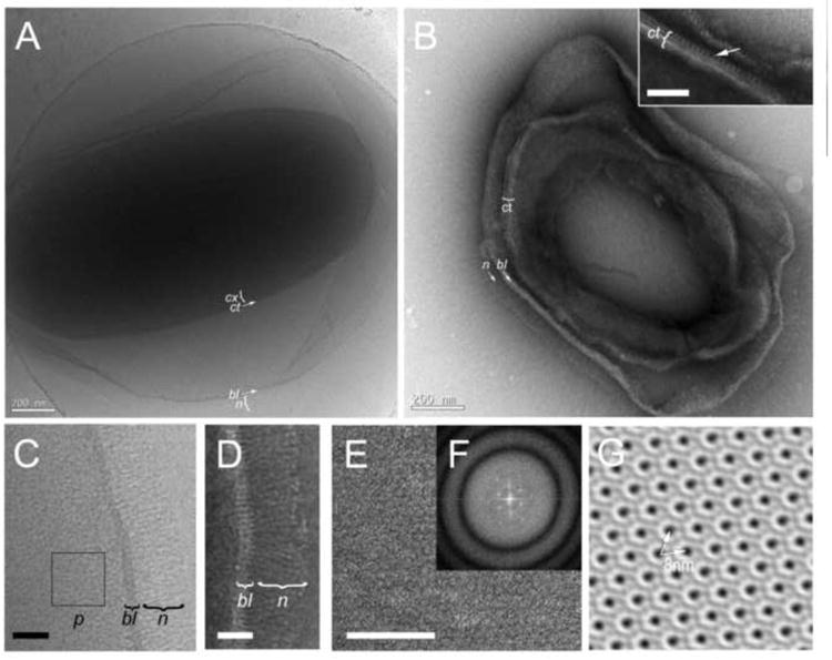
Bacillus anthracis and other pathogenic Bacillus species form spores that are surrounded by an exosporium, a balloon-like layer that acts as the outer permeability barrier of the spore and contributes to spore survival and virulence. The exosporium consists of a hair-like nap and a paracrystalline basal layer. The filaments of the nap are comprised of trimers of the collagen-like glycoprotein BclA, while the basal layer contains approximately 20 different proteins. One of these proteins, BxpB, forms tight complexes with BclA and is required for attachment of essentially all BclA filaments to the basal layer. Another basal layer protein, ExsB, is required for the stable attachment of the exosporium to the spore. To determine the organization of BclA and BxpB within the exosporium, we used cryo-electron microscopy, cryo-sectioning and crystallographic analysis of negatively stained exosporium fragments to compare wildtype spores and mutant spores lacking BclA, BxpB or ExsB (ΔbclA, ΔbxpB and ΔexsB spores, respectively). The trimeric BclA filaments are attached to basal layer surface protrusions that appear to be trimers of BxpB. The protrusions interact with a crystalline layer of hexagonal subunits formed by other basal layer proteins. Although ΔbxpB spores retain the hexagonal subunits, the basal layer is not organized with crystalline order and lacks basal layer protrusions and most BclA filaments, indicating a central role for BxpB in exosporium organization.

Keywords: 2D crystallography; Anthrax spores; Bacterial ultrastructure; Cryo-electron microscopy; Cryo-sectioning; Sporulation; Tokuyasu technique.


Figures

Figure 1
Figure 1
Electron microscopy of the wildtype B. anthracis spore. (A) Cryo-EM of a frozen-hydrated, intact spore. (B) Negatively stained cryo-section (Tokuyasu method). Scale bars are 200 nm. The basal layer (bl), nap (n), coat (ct) and cortex (cx) are indicated. The inset shows a closeup view of the periodic outer layer (arrow) in the coat. Scale bar = 50 nm. (C) Detail of the wildtype exosporium by cryo-EM. The punctate pattern (p) is indicated. (D) Detail of the ultrathin cryo-section, showing the striated basal layer (bl) and nap filaments (n) with terminal knobs. (E) Part of a negatively stained cryo-section showing an exosporium flat in the plane of the grid. The hexagonal paracrystalline pattern is clearly evident. (F) Power spectrum of the Fourier transform of (E), showing diffraction maxima corresponding to the crystal array. Panel (G) is derived from (E) by 2D crystal processing, without the application of symmetry. The 8 nm lattice spacing is indicated (arrows). Scale bars in C, D and E are 50 nm.
Figure 2
Figure 2
Electron microscopy of mutant B. anthracis spores. (A) Cryo-EM and (B) negatively stained, ultrathin cryo-section of the ΔbclA spore. Detail view of the ΔbclA exosporium by cryo-EM (C) and cryo-sectioning (D). The basal layer (bl) and the hexagonal pattern (h) are indicated. (E) Cryo-EM and (F) ultrathin cryo-section of the ΔbxpB spore. Detail of the ΔbxpB exosporium by cryo-EM (G) and cryo-sectioning (H). The coat (ct) and basal layer (bl) are indicated. (I) Cryo-section of the ΔexsB spore. Scale bars in A, B, E and F are 200 nm; in C, D, G and H, 50 nm; and in I, 100 nm.
Figure 3
Figure 3
2D crystallographic analysis of isolated exosporium fragments. (A-C) Micrographs of the negatively stained wildtype (A), ΔbclA (B) and ΔbxpB (C) exosporium and the power spectra of their Fourier transforms (insets). Scale bars are 50 nm. (D-F) Contour plots of the p6 symmetry projection maps of the wildtype, ΔbclA and ΔbxpB exosporium reconstructions are shown in in D, E and F, respectively. Positive density is shown in grayscale, negative density as red, dashed contours. (G-I) Contour plots of the difference maps between the wildtype and ΔbclA exosporium (G), between ΔbclA and ΔbxpB (H) and between the wildtype and ΔbxpB exosporium (I). In the difference maps, green contours are positive, red are negative. All maps were plotted on the same density scale.
Figure 4
Figure 4
Schematic diagram of the organization of BxpB (blue) and BclA (red) trimers in the exosporium. The two layers of density discernible in the 12-14 nm thick basal layer are shown in gray with the side views of the hexagonal subunits superimposed. The black rectangle indicates the hole in the middle of the hexagonal subunits. A magnified image of a wildtype cryo-section is shown below the schematic with relevant features superimposed. The p6 averaged surface view of the exosporium from the 2D reconstruction is shown on the left with the proposed location of the BxpB trimers shown (blue and open circles) on part of the array. Relevant dimensions are indicated. See text for details.

References

    1. Ascenzi P, Visca P, Ippolito G, Spallarosa A, Bolognesi M, Montecucco C. Anthrax toxin: a tripartite lethal combination. FEBS Let. 2002;531:384–388. - PubMed
    1. Ball DA, Taylor R, Todd SJ, Redmond C, Couture-Tosi E, Sylvestre P, Moir A, Bullough PA. Structure of the exosporium and sublayers of spores of the Bacillus cereus family revealed by electron crystallography. Mol Microbiol. 2008;68:947–958. - PubMed
    1. Basu S, Kang TJ, Chen WH, Fenton MJ, Baillie L, Hibbs S, Cross AS. Role of Bacillus anthracis spore structures in macrophage cytokine responses. Infect Immun. 2007;75:2351–2358. - PMC - PubMed
    1. Beaman TC, Pankratz HS, Gerhardt P. Paracrystalline sheets reaggregated from solubilized exosporium of Bacillus cereus. J Bacteriol. 1971;107:320–324. - PMC - PubMed
    1. Beaman TC, Pankratz HS, Gerhardt P. Ultrastructure of the exosporium and underlying inclusions in spores of Bacillus megaterium. J Bacteriol. 1972;109:1198–1209. - PMC - PubMed

Publication types

MeSH terms

Substances