Review
. 2014 Apr:121:178-93.
doi: 10.1016/j.exer.2014.02.007. Epub 2014 Mar 4.

Wounding the cornea to learn how it heals

Affiliations
Review

Wounding the cornea to learn how it heals

Mary Ann Stepp et al. Exp Eye Res. 2014 Apr.

Abstract

Corneal wound healing studies have a long history and rich literature that describes the data obtained over the past 70 years using many different species of animals and methods of injury. These studies have lead to reduced suffering and provided clues to treatments that are now helping patients live more productive lives. In spite of the progress made, further research is required since blindness and reduced quality of life due to corneal scarring still happens. The purpose of this review is to summarize what is known about different types of wound and animal models used to study corneal wound healing. The subject of corneal wound healing is broad and includes chemical and mechanical wound models. This review focuses on mechanical injury models involving debridement and keratectomy wounds to reflect the authors' expertise.

Keywords: animal models; cornea; debridement; keratectomy; mouse strain; wound healing.


Figures

Figure 1
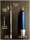
Figure 1. The mouse cornea can be wounded with a dulled blade or rotating burr (AlgerbrushII)
Typically, a 1.5 mm trephine is used to demarcate a circular area at the center of the cornea and the edge of the dulled blade is used to remove epithelial cells. The dulled blade is held by a blade breaker. Alternatively, a roating burr equipped with a 0.5mm burr can be used. After wounding, the epithelial tissue removed can be frozen in liquid nitrogen and used for biochemical or molecular analyses. Trephine sizes can by varied to create smaller (1.0 mm) or larger (2.0 or 2.5 mm) wounds in the mouse. For the rat, 3.0 mm to 4.0 mm trephines are used.
Figure 2
Figure 2. After 1.5 and 2.5 mm debridement wounds made using the dulled blade, the mouse cornea spontaneously develops erosions most often located in the nasal quadrant
The images on top show mouse corneas with a 1.5 or a 2.5 mm debridement wound stained either immediately with the vital dye Richardson stain or allowed to heal for 18 hr or 4 weeks. Note that wounds close rapidly after both wound types and small erosions are present 4 weeks after wounding. To determine the sites where erosions are most likely to form, the corneas of BALB/c mice were wounded and allowed to heal for 4 weeks. At the time of sacrifice, eyes were stained Richardstain stain to reveal exposed basement membrane and the locations of the erosions determined by quadrant. Corneas lacking erosions are considered as healed. Although there was a greater tendancy after large wounds for erosions to be in the center compared to small wounds, more erosions were present at the nasal quardrant of the cornea after 1.5 and 2.5 mm wounds.
Figure 3
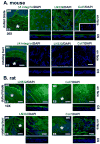
Figure 3. Rotating-burr wounds remove basement membrane components LN332 and type VII collagen at the center of the wounded mouse cornea but leave patches of both proteins behind on the rat cornea
Representative images showing the status of the BMZ immediately after dulled blade and rotating burr wounds in the mouse (A) and rat (B). On the right are en face 63x images of the regions indicated by the asterisks on the 20x (mouse) or 10x (rat) images shown on the left to highlight the localization of two basement membrane components, LN332 and type VII collagen, shown in green and nuclei stained with DAPI shown in blue. For the mouse, the en face 63x confocal images were merged images obtained from 18 confocal layers taken at 1 mm intervals; CS images are 3D reconstructions obtained by rotating 18 layers at a 90° angle to obtain cross sections. For the rat, the en face confocal images were merged images obtained from 40 confocal layers taken at 1 mm intervals; CS images are 3D reconstructions obtained by rotating 40 layers at a 90° angle to obtain cross sections. Note the presence of an intact basement membrane after dulled-blade wounding in both mouse and rat corneas and its disappearance in the mouse and disruption in the rat after rotating-burr wounding. The mouse data shown above were taken from Pal-Ghosh et al., 2011b; the rat data were generated using ithe same methods and antibodies. WE = wound edge; ES = exposed stroma. Bar in 20x image = 50 μm; Bar in 10x image = 100 μm; and bar in 63x image = 7 μm.
Figure 4
Figure 4. Sub-basal nerves and deeper stromal nerves become disassembled within hours after enucleation in organ culture
Shown here are whole mount confocal images obtained after staining mouse corneas with the classIII βtubulin antibody (TuJ1) in red and keratin-8 in green with the central cornea shown above and the limbal region shown below. K8+ goblet cells define the the corneal:limbal junction (Pajoohesh-Ganji et al., 2012). Corneas were either fixed immediately after sacrifice (0 hr) or placed in organ culture for 1, 3, or 6 hrs prior to fixing and staining. A minimum of 3 corneas each were used for these studies and the method and antibodies used for these studies have been described (Pal-Ghosh et al., 2011b). The sub-basal nerves form distinct swirling patterns and appear beaded and varicose. The sites indicated by the white asterisks have been digitally magnified 4-fold and are shown below lower magnification images. The thinnest nerve fibers become less distinct 1 and 3 hrs after corneas are placed in organ culture. By 6 hours the sub-basal nerves are barely detected in the central cornea and only the larger nerves can be seen at the limbus. Bar = 20 μm.

Similar articles

Cited by

References

    1. Acheampong AA, Shackleton M, Tang-Liu DD, Ding S, Stern ME, Decker R. Distribution of cyclosporin A in ocular tissues after topical administration to albino rabbits and beagle dogs. Current eye research. 1999;18:91–103. - PubMed
    1. Adams JM, Harris AW, Pinkert CA, Corcoran LM, Alexander WS, Cory S, Palmiter RD, Brinster RL. The c-myc oncogene driven by immunoglobulin enhancers induces lymphoid malignancy in transgenic mice. Nature. 1985;318:533–538. - PubMed
    1. Amescua G, Collings F, Sidani A, Bonfield TL, Rodriguez JP, Galor A, Medina C, Yang X, Perez VL. Effect of CXCL-1/KC production in high risk vascularized corneal allografts on T cell recruitment and graft rejection. Transplantation. 2008;85:615–625. - PubMed
    1. Anderson RA. Actin filaments in normal and migrating corneal epithelial cells. Investigative ophthalmology & visual science. 1977;16:161–166. - PubMed
    1. Araki-Sasaki K, Ohashi Y, Sasabe T, Hayashi K, Watanabe H, Tano Y, Handa H. An SV40-immortalized human corneal epithelial cell line and its characterization. Investigative ophthalmology & visual science. 1995;36:614–621. - PubMed

Publication types