. 2014 Apr;73(4):295-304.
doi: 10.1097/NEN.0000000000000052.

Mild cognitive impairment and asymptomatic Alzheimer disease subjects: equivalent β-amyloid and tau loads with divergent cognitive outcomes

Affiliations

Mild cognitive impairment and asymptomatic Alzheimer disease subjects: equivalent β-amyloid and tau loads with divergent cognitive outcomes

Diego Iacono et al. J Neuropathol Exp Neurol. 2014 Apr.

Abstract

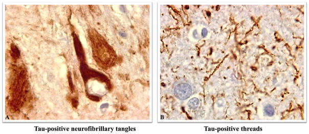
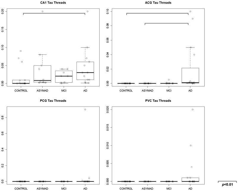
Older adults with intact cognition before death and substantial Alzheimer disease (AD) lesions at autopsy have been termed "asymptomatic AD subjects" (ASYMAD). We previously reported hypertrophy of neuronal cell bodies, nuclei, and nucleoli in the CA1 of the hippocampus (CA1), anterior cingulate gyrus, posterior cingulate gyrus, and primary visual cortex of ASYMAD versus age-matched Control and mild cognitive impairment (MCI) subjects. However, it was unclear whether the neuronal hypertrophy could be attributed to differences in the severity of AD pathology. Here, we performed quantitative analyses of the severity of β-amyloid (Aβ) and phosphorylated tau (tau) loads in the brains of ASYMAD, Control, MCI, and AD subjects (n = 15 per group) from the Baltimore Longitudinal Study of Aging. Tissue sections from CA1, anterior cingulate gyrus, posterior cingulate gyrus, and primary visual cortex were immunostained for Aβ and tau; the respective loads were assessed using unbiased stereology by measuring the fractional areas of immunoreactivity for each protein in each region. The ASYMAD and MCI groups did not differ in Aβ and tau loads. These data confirm that ASYMAD and MCI subjects have comparable loads of insoluble Aβ and tau in regions vulnerable to AD pathology despite divergent cognitive outcomes. These findings imply that cognitive impairment in AD may be caused or modulated by factors other than insoluble forms of Aβ and tau.


Figures

FIGURE 1
FIGURE 1
Different types of β-amyloid (Aβ) deposits in the human cortex. The Aβ deposition was detected with the 6E10 antibody, which recognizes predominantly the 1-42Aβ form. (A, B) Diffuse-Aβ. (C, D) Dense-Aβ. All panels were photographed with 20 × objective.
FIGURE 2
FIGURE 2
Different types of tau deposits in the human cortex. Tau deposition was detected with the PHF-1 antibody. (A) Tau-positive neurofibrillary tangles. (B) Tau-positive threads. Panels were photographed with a 40× objective.
FIGURE 3
FIGURE 3
Diffuse β-amyloid (dif-Aβ) in cortical regions. Box plots show the estimated quantities of dif-Aβ in each cortical region (CA1 of the hippocampus [CA1], anterior cingulate gyrus [ACG], posterior cingulate gyrus [PCG], primary visual cortex [PVC]) across all groups in the study (Controls, asymptomatic Alzheimer disease [ASYMAD], mild cognitive impairment [MCI], Alzheimer disease [AD]). Area fraction values (percentages of the 6E10-positive area fraction) are represented on the y axis. The line in the middle of each box represents the median; upper and lower horizontal lines represent SDs. Each spot represents a single measurement. The significance across groups (p < 0.01) when present is indicated by lines between groups.
FIGURE 4
FIGURE 4
Dense β-amyloid (Aβ) in cortical regions. Box plots showing the estimated quantities of Aβ in each cortical region (CA1 of the hippocampus [CA1], anterior cingulate gyrus [ACG], posterior cingulate gyrus [PCG], primary visual cortex [PVC]) across all groups in the study (Controls, asymptomatic Alzheimer disease [ASYMAD], mild cognitive impairment [MCI], Alzheimer disease [AD]). Area fraction values (percentage of the 6E10-positive area fraction) are indicated on the y axis. The line in the middle of each box represents the median; upper and lower horizontal lines represent SDs. Each spot represents a single measurement. Significance across groups (p < 0.01) when present is indicated by lines between groups.
FIGURE 5
FIGURE 5
Tau-positive neurofibrillary tangles (Tau-NFTs) in cortical regions. Box plots showing the estimated quantities of Tau-NFT in each cortical region (CA1 of the hippocampus [CA1], anterior cingulate gyrus [ACG], posterior cingulate gyrus [PCG], primary visual cortex [PVC]) across all groups in the study (Controls, asymptomatic Alzheimer disease [ASYMAD], mild cognitive impairment [MCI], Alzheimer disease [AD]). Each box represents area fraction values (percentage of the PHF-1–positive area fraction). The line in the middle of each box represents the median; upper and lower horizontal lines represent the SDs. Each spot represents a single measurement. Significance across groups (p < 0.01) when present is indicated by lines between groups.
FIGURE 6
FIGURE 6
Tau-positive neuropil threads (Tau-th) in cortical regions. Box plots showing the estimated quantities of Tau-th in each cortical region (CA1 of the hippocampus [CA1], anterior cingulate gyrus [ACG], posterior cingulate gyrus [PCG], primary visual cortex [PVC]) across all groups in the study (Controls, asymptomatic Alzheimer disease [ASYMAD], mild cognitive impairment [MCI], Alzheimer disease [AD]). Each box represents the area fraction value (percentage of the PHF-1–positive area fraction). Lines in the middle of a box represent the median; upper and lower horizontal lines represent SDs. Each spot represents a single measurement. Significance across groups (p < 0.01) when present is indicated by lines between groups.

References

    1. Tomlinson BE, Blessed G, Roth M. Observations on the brains of nondemented old people. J Neurol Sci. 1968;7:331–56. - PubMed
    1. Katzman R, Terry R, DeTeresa R, et al. Clinical, pathological, and neurochemical changes in dementia: A subgroup with preserved mental status and numerous neocortical plaques. Ann Neurol. 1988;23:138–44. - PubMed
    1. Crystal H, Dickson D, Fuld P, et al. Clinicopathologic studies in dementia: Nondemented subjects with pathologically confirmed Alzheimer’s disease. Neurology. 1988;38:1682–87. - PubMed
    1. Knopman DS, Parisi JE, Salviati A, et al. Neuropathology of cognitively normal elderly. J Neuropathol Exp Neurol. 2003;62:1087–95. - PubMed
    1. Troncoso JC, Martin LJ, Dal Forno G, et al. Neuropathology in controls and demented subjects from the Baltimore Longitudinal Study of Aging. Neurobiol Aging. 1996;17:365–71. - PubMed

Publication types