

Prion protein-specific antibodies that detect multiple TSE agents with high sensitivity
- PMID: 24608105
- PMCID: PMC3946747
- DOI: 10.1371/journal.pone.0091143
Prion protein-specific antibodies that detect multiple TSE agents with high sensitivity
Abstract
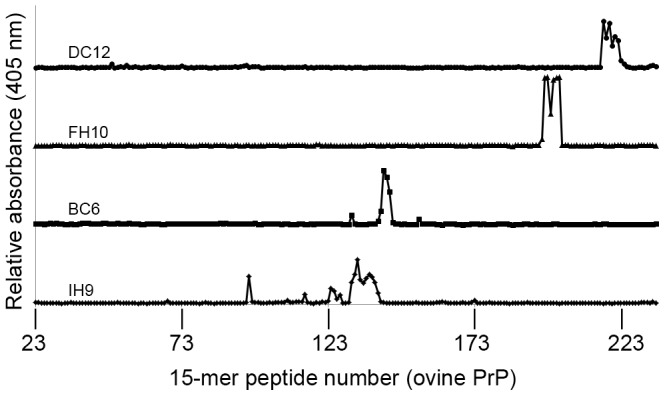
This paper describes the generation, characterisation and potential applications of a panel of novel anti-prion protein monoclonal antibodies (mAbs). The mAbs were generated by immunising PRNP null mice, using a variety of regimes, with a truncated form of recombinant ovine prion protein spanning residues 94-233. Epitopes of specific antibodies were mapped using solid-phase Pepscan analysis and clustered to four distinct regions within the PrP molecule. We have demonstrated the utility of these antibodies by use of Western blotting and immunohistochemistry in tissues from a range of different species affected by transmissible spongiform encephalopathy (TSE). In comparative tests against extensively-used and widely-published, commercially available antibodies, similar or improved results can be obtained using these new mAbs, specifically in terms of sensitivity of detection. Since many of these antibodies recognise native PrPC, they could also be applied to a broad range of immunoassays such as flow cytometry, DELFIA analysis or immunoprecipitation. We are using these reagents to increase our understanding of TSE pathogenesis and for use in potential diagnostic screening assays.
Conflict of interest statement
Figures


Similar articles
-
Species-specificity of a panel of prion protein antibodies for the immunohistochemical study of animal and human prion diseases.J Comp Pathol. 2007 Jan;136(1):9-17. doi: 10.1016/j.jcpa.2006.09.002. Epub 2007 Jan 31. J Comp Pathol. 2007. PMID: 17270205
-
Antigenic features of prion proteins of sheep and of other mammalian species.J Immunol Methods. 1997 Aug 22;207(1):89-101. doi: 10.1016/s0022-1759(97)00121-x. J Immunol Methods. 1997. PMID: 9328590
-
Generation of antibodies against bovine recombinant prion protein in various strains of mice.Clin Vaccine Immunol. 2006 Jan;13(1):98-105. doi: 10.1128/CVI.13.1.98-105.2006. Clin Vaccine Immunol. 2006. PMID: 16426006 Free PMC article.
-
The role of antibodies to PrP in the diagnosis of transmissible spongiform encephalopathies.Dev Biol Stand. 1993;80:141-51. Dev Biol Stand. 1993. PMID: 8270104 Review.
-
Vaccine approaches to prevent and treat prion infection : progress and challenges.BioDrugs. 2008;22(1):45-52. doi: 10.2165/00063030-200822010-00005. BioDrugs. 2008. PMID: 18215090 Review.
Cited by
-
Innate Immune Tolerance in Microglia Does Not Impact on Central Nervous System Prion Disease.Front Cell Neurosci. 2022 Jun 30;16:918883. doi: 10.3389/fncel.2022.918883. eCollection 2022. Front Cell Neurosci. 2022. PMID: 35875357 Free PMC article.
-
Caprine PRNP polymorphisms N146S and Q222K are associated with proteolytic cleavage of PrPC.Genet Sel Evol. 2021 Jun 19;53(1):52. doi: 10.1186/s12711-021-00646-x. Genet Sel Evol. 2021. PMID: 34147084 Free PMC article.
-
PrP aggregation can be seeded by pre-formed recombinant PrP amyloid fibrils without the replication of infectious prions.Acta Neuropathol. 2016 Oct;132(4):611-24. doi: 10.1007/s00401-016-1594-5. Epub 2016 Jul 4. Acta Neuropathol. 2016. PMID: 27376534 Free PMC article.
-
Spermine increases acetylation of tubulins and facilitates autophagic degradation of prion aggregates.Sci Rep. 2018 Jul 3;8(1):10004. doi: 10.1038/s41598-018-28296-y. Sci Rep. 2018. PMID: 29968775 Free PMC article.
-
Preclinical transmission of prions by blood transfusion is influenced by donor genotype and route of infection.PLoS Pathog. 2021 Feb 18;17(2):e1009276. doi: 10.1371/journal.ppat.1009276. eCollection 2021 Feb. PLoS Pathog. 2021. PMID: 33600501 Free PMC article.
References
-
- Gretzschel A, Buschmann A, Langeveld J, Groschup MH (2006) Immunological characterization of abnormal prion protein from atypical scrapie cases in sheep using a panel of monoclonal antibodies. J Gen Virol 87: 3715–3722. - PubMed
-
- Benestad SL, Sarradin P, Thu B, Schonheit J, Tranulis MA, et al. (2003) Cases of scrapie with unusual features in Norway and designation of a new type, Nor98. Vet Rec 153: 202–208. - PubMed
-
- Jeffrey M, Martin S, Gonzalez L (2003) Cell-associated variants of disease-specific prion protein immunolabelling are found in different sources of sheep transmissible spongiform encephalopathy. J Gen Virol 84: 1033–1045. - PubMed
-
- Jeffrey M, Martin S, Gonzalez L, Ryder SJ, Bellworthy SJ, et al. (2001) Differential diagnosis of infections with the bovine spongiform encephalopathy (BSE) and scrapie agents in sheep. J Comp Pathol 125: 271–284. - PubMed
Publication types
MeSH terms
Substances
Grants and funding
LinkOut - more resources
Full Text Sources
Other Literature Sources
Research Materials
