Review
. 2014 Apr;12(4):274-88.
doi: 10.1038/nrmicro3235. Epub 2014 Mar 10.

Bordetella pertussis pathogenesis: current and future challenges

Affiliations
Review

Bordetella pertussis pathogenesis: current and future challenges

Jeffrey A Melvin et al. Nat Rev Microbiol. 2014 Apr.

Abstract

Pertussis, also known as whooping cough, has recently re-emerged as a major public health threat despite high levels of vaccination against the aetiological agent Bordetella pertussis. In this Review, we describe the pathogenesis of this disease, with a focus on recent mechanistic insights into B. pertussis virulence-factor function. We also discuss the changing epidemiology of pertussis and the challenges facing vaccine development. Despite decades of research, many aspects of B. pertussis physiology and pathogenesis remain poorly understood. We highlight knowledge gaps that must be addressed to develop improved vaccines and therapeutic strategies.


Conflict of interest statement

Competing interests statement

The authors declare no competing interests.

Figures
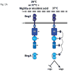
Figure 1
Figure 1. The BvgAS master regulatory system
(a) BvgS is a polydomain histidine sensor kinase containing (from the N- to the C-terminus) two periplasmically-located venus flytrap domains (VFT1 & VFT2), a transmembrane domain, a PAS domain (PAS), a histidine kinase domain (HK), a receiver domain (Rec) and a histidine phosphoryl transfer domain (Hpt). BvgA is a response regulator protein with an N-terminal receiver domain (Rec) and a C-terminal helix-turn-helix domain (HTH). BvgS is activate at 37°C, autophosphorylates at a conserved histidine in the HK domain, and transfers the phosphoryl group to the Rec, the Hpt and then to the Rec domain of BvgA. Phosphorylated BvgA (BvgA-P) activates expression of virulence-associated genes (vags; which are subdivided into class 1 and 2 genes) and represses expression of virulence-repressed genes (vrgs; known as class 4 genes). BvgS is inactive and remains unphosphorylated when bacteria are grown at a low temperature (~25°C) or at 37°C in the presence of chemical modulators (such as MgSO4 or nicotinic acid). (OM-outer membrane, CM-cytoplasmic membrane) (b) BvgAS controls four classes of genes and three distinct phenotypic phases. The Bvg+ phase occurs when BvgAS is fully active and is characterized by maximal expression of genes encoding adhesins (class 2 genes, such as fhaB, fim2 and fim3, expression levels indicated by an orange line) and toxins (class 1 genes, such as cyaA-E, ptx-ptl and bsc genes, expression levels indicated by a red line), and minimal expression of class 3 and class 4 genes (expression levels indicated by purple and blue lines, respectively). The Bvg+ phase is necessary and sufficient to cause respiratory infection (i.e., in vivo). The Bvg phase occurs when BvgAS is inactive and is characterized by maximal expression of class 4 genes and minimal expression of class 1, 2, and 3 genes. (Note that regulation of some vrgs is indirect; when BvgAS is inactive, it does not repress frlAB, a positive regulator at the top of the motility regulon, and it does not activate bvgR, a negative regulator of vrg loci.) The Bvg phase is required for growth under nutrient limiting conditions, such as may be encountered in the environment (i.e., ex vivo). The Bvgi phase occurs when BvgAS is partially active and is characterized by maximal expression of class 3 genes and minimal expression of class 1, 2, and 4 genes. The only class 3 gene characterized so far is bipA, which is activated by BvgA under Bvgi phase conditions and repressed by BvgA under Bvg+ phase conditions. The Bvgi phase may be important for transmission between hosts, but this has not been fully elucidated.
Figure 2
Figure 2. Toxin-mediated virulence of Bordetella spp
(a) Pertussis toxin (PT, PDB ID 1PRT), is an AB5-type toxin composed of one catalytic subunit (A subunit) and five membrane-binding/transport subunits (B subunits). PT is assembled in the bacterial periplasm and exported by a type IV secretion system. (b) On binding to a sialoglycoprotein host cell receptor, PT is endocytosed and trafficked through the Golgi to the endoplasmic reticulum. In the endoplasmic reticulum, the B5 complex binds to ATP and dissociates from the A subunit. The A subunit is then transported into the cytoplasm and traffics on exosomes to the cytoplasmic membrane, where it ADP-ribosylates the α subunit of heterotrimeric G proteins. This modification alters the ability of G proteins to regulate multiple enzymes and pathways, including their ability to inhibit cyclic AMP (cAMP) formation. The overall result of these modifications is an initial suppression of inflammatory cytokine production and inhibition of immune cell recruitment to the site of infection. (c) Bordetella spp. adenylate cyclase toxin (ACT) is composed of two primary domains, a calmodulin-responsive adenylate cyclase enzymatic domain (yellow) and an RTX domain (black), which are connected by hydrophobic segments (green). (d) The RTX domain of ACT interacts with CR3 receptors that are expressed on host cell membranes from a wide range of cell types. The hydrophobic segments of the linker region (green) form pores in the membrane that enable the passage of ions and translocation of the adenylate cyclase domain into the cytoplasm. Adenylate cyclase activity is stimulated by binding to calmodulin in the host cell. The combined effects of ACT intoxication and pore formation result in inhibition of complement-dependent phagocytosis, induction of anti-inflammatory cytokines, suppression of pro-inflammatory cytokines and inhibition of immune cell recruitment.
Figure 3
Figure 3. Presentation of filamentous hemagglutinin, fimbriae and pertactin on the Bordetella cell surface
(a) Filamentous hemagglutinin (FHA) is a TpsA exoprotein (blue) that is translocated across the outer membrane through its cognate TpsB pore protein (red), FhaC. This translocation occurs via the two-partner secretion pathway. Processing during translocation removes the C-terminal prodomain (yellow) from the full-length FhaB protein to produce the mature ~250 kDa FHA protein. FHA is required for adherence to ciliated epithelial cells and for persistence during infection, possibly by directly or indirectly modulating the host immune system. (b) Bordetella spp. fimbriae are type 1 pili. FimB is similar to chaperone proteins that traffic major fimbrial subunits (Fim2 and Fim3, in this case) to the membrane usher FimC. FimB and FimC are necessary for fimbrial secretion and FimD (the tip subunit) is necessary for fimbrial assembly. Fimbriae are required for persistence during infection, possibly by functioning similarly to FHA by directly or indirectly modulating the immune system. Furthermore, studies have suggested that fimbriae are necessary for adherence to ciliated epithelial cells. (c) Pertactin is a classical autotransporter. The C-terminal ~30 kDa region (red) forms a channel in the outer membrane (om) that is required for translocation of the ~70 kDa β-helical passenger domain (blue) to the cell surface. Although the precise role of pertactin is unclear, data suggests that pertactin may contribute to virulence by resisting neutrophil-mediated clearance.

References

    1. de Greeff SC, et al. Pertussis disease burden in the household: how to protect young infants. Clin Infect Dis. 2010;50:1339–45. - PubMed
    1. Bordet J, Gengou O. Le microbe de la coqueluche. Annales de I’Institut Pasteur. 1906;20:731–741.
    1. Cherry JD. Why do pertussis vaccines fail? Pediatrics. 2012;129:968–70. - PubMed
    1. Diavatopoulos DA, et al. Bordetella pertussis, the causative agent of whooping cough, evolved from a distinct, human-associated lineage of B. bronchiseptica. PLoS Pathog. 2005;1:e45. This study identified four complexes of Bordetella and suggested that B. pertussis and B. parapertussis independently evolved from a B. bronchiseptica-like ancestor. - PMC - PubMed
    1. Park J, et al. Comparative genomics of the classical Bordetella subspecies: the evolution and exchange of virulence-associated diversity amongst closely related pathogens. BMC Genomics. 2012;13:545. - PMC - PubMed

Publication types

MeSH terms