. 2015 Feb 12;34(7):846-56.
doi: 10.1038/onc.2014.27. Epub 2014 Mar 10.

Mdmx promotes genomic instability independent of p53 and Mdm2

Affiliations

Mdmx promotes genomic instability independent of p53 and Mdm2

A M Carrillo et al. Oncogene. .

Abstract

The oncogene Mdmx is overexpressed in many human malignancies, and together with Mdm2, negatively regulates the p53 tumor suppressor. However, a p53-independent function of Mdmx that impacts genome stability has been described, but this function is not well understood. In the present study, we determined that of the 13 different cancer types evaluated, 6-90% of those that had elevated levels of Mdmx had concurrent inactivation (mutated or deleted) of p53. We show elevated levels of Mdmx-inhibited double-strand DNA break repair and induced chromosome and chromatid breaks independent of p53, leading to genome instability. Mdmx impaired early DNA damage-response signaling, such as phosphorylation of the serine/threonine-glutamine motif, mediated by the ATM kinase. Moreover, we identified Mdmx associated with Nbs1 of the Mre11-Rad50-Nbs1 (MRN) DNA repair complex, and this association increased upon DNA damage and was detected at chromatin. Elevated Mdmx levels also increased cellular transformation in a p53-independent manner. Unexpectedly, all Mdmx-mediated phenotypes also occurred in cells lacking Mdm2 and were independent of the Mdm2-binding domain (RING) of Mdmx. Therefore, Mdmx-mediated inhibition of the DNA damage response resulted in delayed DNA repair and increased genome instability and transformation independent of p53 and Mdm2. Our results reveal a novel p53- and Mdm2-independent oncogenic function of Mdmx that provides new insight into the many cancers that overexpress Mdmx.


Conflict of interest statement

Conflict of interest

The authors declare no potential conflict of interest.

Figures

Figure 1
Figure 1. Elevated Mdmx levels promote genome instability independent of p53
p53−/− MEFs were infected with a bicistronic retrovirus encoding YFP alone (Vector; n=96), YFP and HA-Mdmx (n=98), or YFP and HA-Mdmx1–345 (MxΔRING; n=119). A) Metaphases were evaluated for chromosome aberrations, and the percentage of cells with the indicated aberration is graphed. Statistical significance was determined using the Fisher’s exact test. B) Representative pictures of a chromatid (left) and a chromosome (right) break in p53−/− MEFs overexpressing Mdmx. Inset is an expanded view of the aberration marked by the arrow.
Figure 2
Figure 2. Mdmx overexpression inhibits double-strand DNA break repair independent of p53 and Mdm2
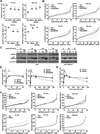
p53−/− (A,B), Arf−/− (C), or p53−/−Mdm2−/− (D,E) MEFs were infected with a bicistronic retrovirus encoding YFP alone (Vector), GFP and HA-Mdm2, YFP and FLAG-Mdmx, YFP and HA-Mdmx, or YFP and HA-MxΔRING. Western blots of the indicated proteins were performed. Following 5 Gy of γIR, neutral comet assays were performed at intervals. All data are a mean of a minimum of three independent experiments. Each symbol in B, C, and E is the mean of an individual experiment and the line is the mean of all experiments. Error bars represent SEM. *p ≤ 0.01 in A; *p < 0.05 in D; each compared to vector control; student’s t-test.
Figure 3
Figure 3. Elevated Mdmx impairs the early DNA damage response signal
p53−/− (A–D, F) or p53−/−Mdm2−/− (A–C, E, F) MEFs were infected with a bicistronic retrovirus encoding YFP alone (vector, V), YFP and full-length HA-Mdmx (Mx) or YFP and HA-MxΔRING (MxΔR). A, B, D–F) Following exposure to 5 Gy of γIR, MEFs were fixed at the indicated intervals and immunofluorescence for γH2AX (A, B) or pS/T-Q (D–F) was performed. The number of foci per cell was quantified. (A, D, E) The mean of at least three independent experiments is graphed. Error bars represent SEM, and significance determined using a confidence interval of 95%. (B, F) Ranking of γH2AX (B) and pS/T-Q (F) foci in individual cells was graphed for a representative experiment in both MEF genotypes. C) Western blot analysis of whole cell lysates for the proteins indicated to the left of the panels at the indicated intervals following γIR.
Figure 4
Figure 4. Mdmx associates with Nbs1 independent of Mdm2
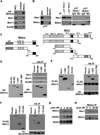
A) Whole cell lysates (WCL) from HA-Mdmx expressing 293T cells were immunoprecipitated (IP) with anti-HA or isotype control antibody and Western blotted. B) Whole cell lysates from p53−/−, p53−/−Mdm2−/−, or p53−/−Mdmx−/− were immunoprecipitated with anti-Mdmx or isotype control antibody and Western blotted. C) Schematic representation of full-length Mdmx and Nbs1 and deletion mutants of each. Binding domains for p53, Mdm2, Mre11, and the acidic, Zinc (Zn), RING, forkhead-associated (FHA), and BRCT domains, and the nuclear localization sequences (NLS) are shown. D–F) 293T cells transfected with empty vector or vectors encoding protein tagged full-length or deletion mutants of Mdmx or Nbs1 as indicated. Whole cell lysates (left panels) and anti-HA or anti-FLAG immunoprecipitations (right panels) were Western blotted. Asterisk denotes immunoglobulin heavy chain and non-specific band in E and F, respectively. G) At intervals (minutes) after 5 Gy of γIR, Western blots were performed on the chromatin-bound protein fraction of p53−/− MEFs. H) Following 5 Gy of γIR for the indicated intervals (minutes), the chromatin-bound protein fraction of p53−/−Mdm2−/− MEFs was immunoprecipitated with anti-Mdmx and Western blots were performed.
Figure 5
Figure 5. Mdmx promotes chromosome instability independent of Mdm2
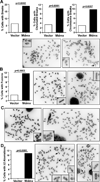
Metaphases from vector control (n=176) or Mdmx overexpressing (n=246) p53−/−Mdm2−/− MEFs were evaluated for chromosome aberrations. A) The percentage of cells with one or more breaks of either kind (chromatid or chromosome) or that have the specified break is graphed. Representative pictures of a chromatid (left) and a chromosome (right) break in p53−/−Mdm2−/− MEFs overexpressing Mdmx are shown. B) The percentage of cells with one or more chromosome fusions is graphed. Representative pictures of a centromere-centromere (left) and a telomere-telomere (right) fusion in p53−/−Mdm2−/− MEFs overexpressing Mdmx are shown. C) Representative pictures of structural aberrations distinct from breaks and fusions detected in p53−/−Mdm2−/− MEFs overexpressing Mdmx are shown. D) The percentage of cells with more than one chromosomal aberration is graphed. Representative pictures of metaphases with multiple aberrations are shown. Insets are expanded views of each aberration marked by an arrow (A–D). Statistical significance was determined using the Fisher’s exact test (A, B, D)
Figure 6
Figure 6. Increased levels of Mdmx promote transformation independent of p53 or Mdm2
Soft agar assays of p53−/− (A) or p53−/−Mdm2−/− (B) MEFs infected with bicistronic retrovirus expressing YFP alone, HA-tagged full-length Mdmx and YFP, or HA-tagged MxΔRING and YFP were performed. Each graph represents the average colonies per well from at least 2 independent experiments with each experiment consisting of three replicates. *p<0.0001 student’s t-test.

References

    1. Momand J, Jung D, Wilczynski S, Niland J. The MDM2 gene amplification database. Nucleic Acids Res. 1998;26:3453–3459. - PMC - PubMed
    1. Rayburn E, Zhang R, He J, Wang H. MDM2 and human malignancies: expression, clinical pathology, prognostic markers, and implications for chemotherapy. Curr Cancer Drug Targets. 2005;5:27–41. - PubMed
    1. Wade M, Li YC, Wahl GM. MDM2, MDMX and p53 in oncogenesis and cancer therapy. Nat Rev Cancer. 2013;13:83–96. - PMC - PubMed
    1. Lundgren K, Montes de Oca Luna R, McNeill YB, Emerick EP, Spencer B, Barfield CR, et al. Targeted expression of MDM2 uncouples S phase from mitosis and inhibits mammary gland development independent of p53. Genes Dev. 1997;11:714–725. - PubMed
    1. Alt JR, Bouska A, Fernandez MR, Cerny RL, Xiao H, Eischen CM. Mdm2 binds to Nbs1 at sites of DNA damage and regulates double strand break repair. J Biol Chem. 2005;280:18771–18781. - PubMed

Publication types

MeSH terms

Substances