. 2014 Mar 7;9(3):e91008.
doi: 10.1371/journal.pone.0091008. eCollection 2014.

Overlapping neural systems represent cognitive effort and reward anticipation

Affiliations

Overlapping neural systems represent cognitive effort and reward anticipation

Eliana Vassena et al. PLoS One. .

Abstract

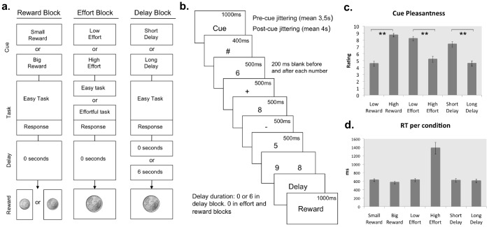
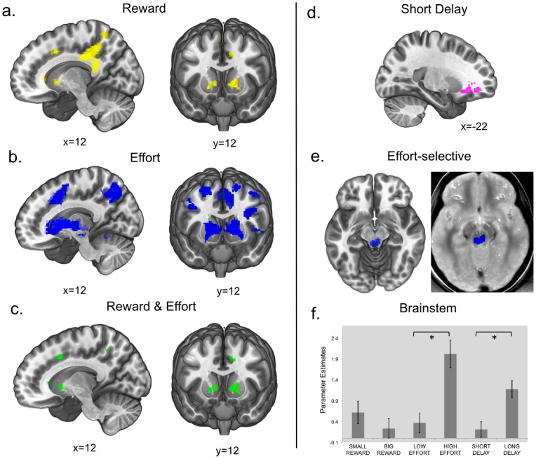
Anticipating a potential benefit and how difficult it will be to obtain it are valuable skills in a constantly changing environment. In the human brain, the anticipation of reward is encoded by the Anterior Cingulate Cortex (ACC) and Striatum. Naturally, potential rewards have an incentive quality, resulting in a motivational effect improving performance. Recently it has been proposed that an upcoming task requiring effort induces a similar anticipation mechanism as reward, relying on the same cortico-limbic network. However, this overlapping anticipatory activity for reward and effort has only been investigated in a perceptual task. Whether this generalizes to high-level cognitive tasks remains to be investigated. To this end, an fMRI experiment was designed to investigate anticipation of reward and effort in cognitive tasks. A mental arithmetic task was implemented, manipulating effort (difficulty), reward, and delay in reward delivery to control for temporal confounds. The goal was to test for the motivational effect induced by the expectation of bigger reward and higher effort. The results showed that the activation elicited by an upcoming difficult task overlapped with higher reward prospect in the ACC and in the striatum, thus highlighting a pivotal role of this circuit in sustaining motivated behavior.


Conflict of interest statement

Competing Interests: The authors have declared that no competing interests exist.

Figures

Figure 1
Figure 1. Task structure and behavioral performance.
a. Block types. In every block only one trial type is presented, where only one feature is manipulated. In a trial in the reward block, the cue informs about the final reward being small or big. In a trial in the effort block, the cue informs about the difficulty level (low or high). In the delay block, the cue informs about the length of the delay between response and reward delivery (short or long). b. Task structure and timing. The cue presentation is followed by a fixation symbol. The task follows, consisting of an addition followed by a subtraction. Two possible results are presented and the subject has to choose the correct one. After the response, a delay can occur. If the response was accurate, the reward is shown. c. Average rating of pleasantness for every cue-type (small reward cue, big reward cue, low effort cue, high effort cue, short delay cue, long delay cue). e. Average reaction times (RTs) in every condition (small reward, big reward, low effort, high effort, short delay, long delay). RT in the high effort condition is significantly higher than in the low effort condition (p<0.001).
Figure 2
Figure 2. fMRI Results.
a. Reward contrast (big reward>small reward). Activation clusters are located in the posterior cingulate cortex (PCC), anterior cingulate cortex (ACC), striatum and superior frontal gyrus (SFG). b. Effort contrast (high effort>low effort). Activation clusters are located in the posterior cingulate cortex (PCC), brainstem, anterior cingulate cortex (ACC), insula, striatum and middle frontal gyrus (MFG). c. Conjunction of high effort>low effort & big reward>small reward. Overlapping activation clusters are located in the striatum, anterior cingulate cortex (ACC) and precuneus. d. Short delay>long delay contrast. The activation cluster is located in the left orbitofrontal cortex (OFC). e. Effort-selective activation ((high effort>low effort)>(high reward> low reward)), SVC for the region of the brainstem, p value 0.05 FWE correction for multiple comparisons, plotted on Proton Density Weighted MRI Template (left image). f. Parameter estimates plot at voxel −4, −32, −10 (MNI coordinates), local maximum in the activation cluster located in the Brainstem in the effort-selective contrast.

References

    1. Rangel A, Camerer C, Montague PR (2008) A framework for studying the neurobiology of value-based decision making. Nat Rev Neurosci 9: 545–556. - PMC - PubMed
    1. Kahneman D, Tversky A (1979) Prospect theory: an analysis of decision under risk. Econometrica 47: 263–292.
    1. Schultz W, Dickinson A (2000) Neuronal coding of prediction errors. Annu Rev Neurosci 23: 473–500. - PubMed
    1. Silvetti M, Seurinck R, Verguts T (2012) Value and prediction error estimation account for volatility effects in ACC: A model-based fMRI study. Cortex 49: 1327–1635. - PubMed
    1. Knutson B, Cooper JC (2005) Functional magnetic resonance imaging of reward prediction. Curr Opin Neurol 18: 411–417. - PubMed

Publication types

LinkOut - more resources