. 2014 Mar 7;9(3):e91072.
doi: 10.1371/journal.pone.0091072. eCollection 2014.

SUMOylation of Myc-family proteins

Affiliations

SUMOylation of Myc-family proteins

Arianna Sabò et al. PLoS One. .

Abstract

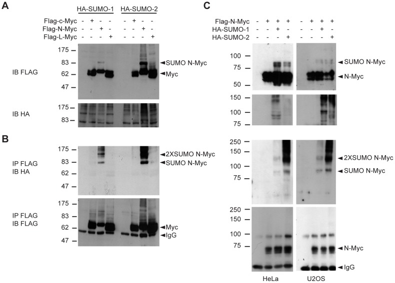
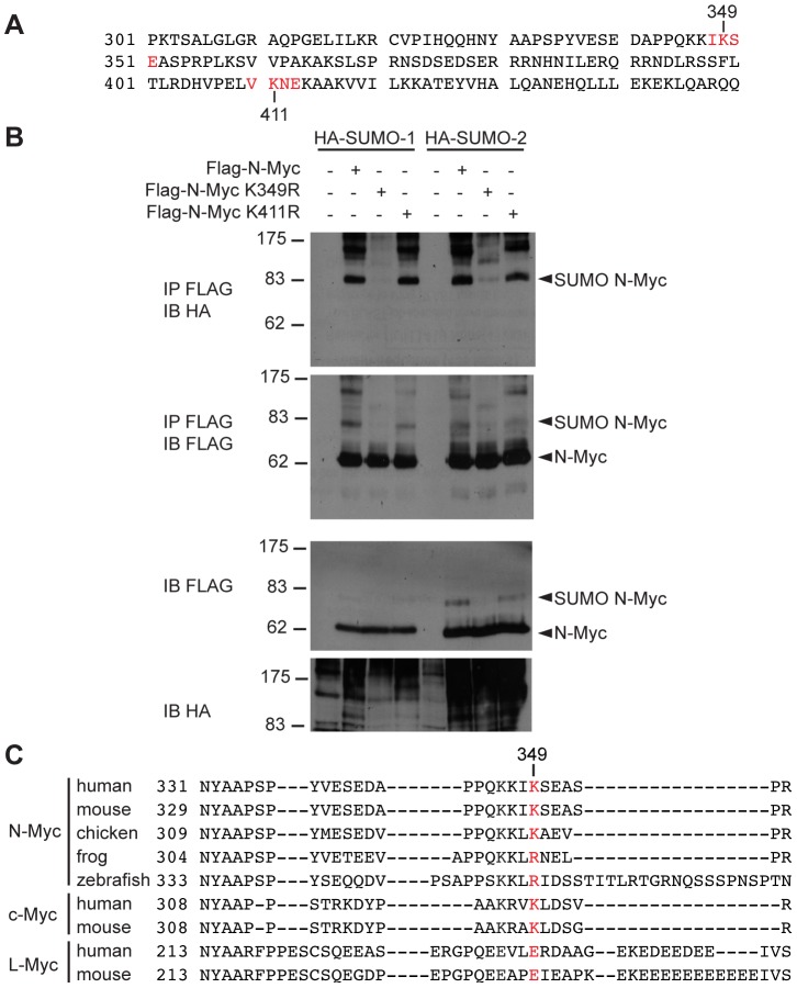
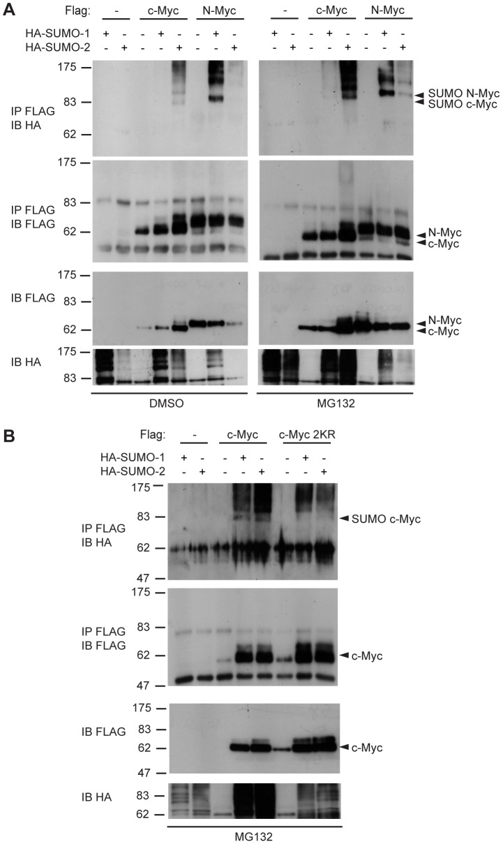
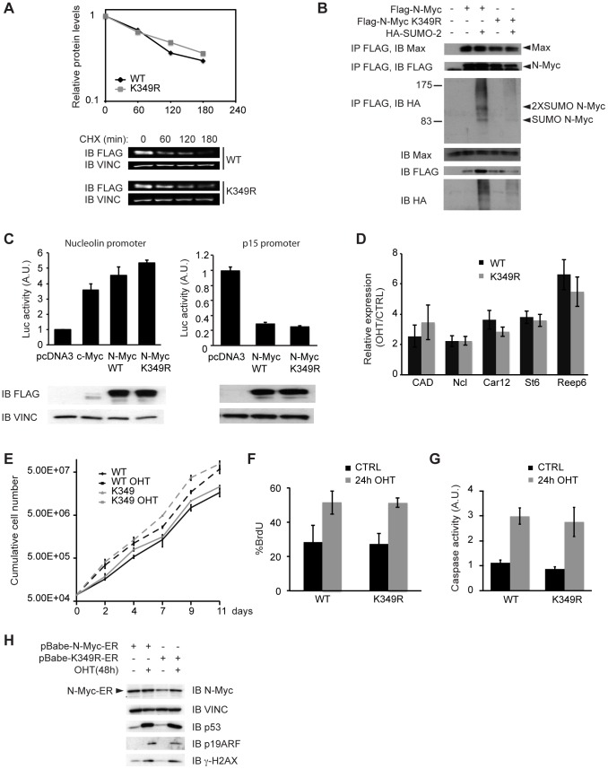
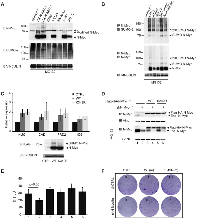
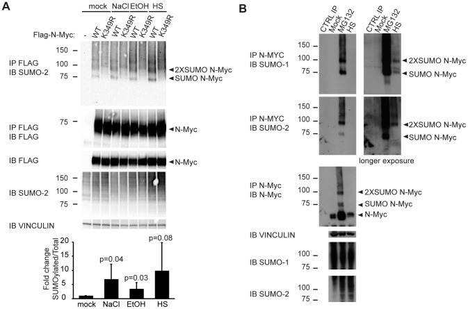
Myc-family proteins are key controllers of the metabolic and proliferative status of the cell, and are subjected to a complex network of regulatory events that guarantee their efficient and fast modulation by extracellular stimuli. Hence, unbalances in regulatory mechanisms leading to altered Myc levels or activities are often reported in cancer cells. Here we show that c- and N-Myc are conjugated to SUMO proteins at conserved lysines in their C-terminal domain. No obvious effects of SUMOylation were detected on bulk N-Myc stability or activities, including the regulation of transcription, proliferation or apoptosis. N-Myc SUMOylation could be induced by cellular stresses, such as heat shock and proteasome inhibition, and in all instances concerned a small fraction of the N-Myc protein. We surmise that, as shown for other substrates, SUMOylation may be part of a quality-control mechanism acting on misfolded Myc proteins.


Conflict of interest statement

Competing Interests: The authors have declared that no competing interests exist.

Figures

Figure 1
Figure 1. SUMOylation of Myc proteins.
(A) Immunoblot analysis in denaturing conditions of 293T cells transfected with plasmids expressing the indicated proteins. (B) Lysates from (A) were immunoprecipitated (IP) in denaturing conditions with anti-Flag beads, and the precipitates subsequently analyzed by immunoblotting (IB) with the indicated antibodies. (C) as in (A) and (B) but for HeLa and U2OS cells.
Figure 2
Figure 2. N-Myc is SUMOylated at lysine 349.
(A) Mouse N-Myc protein sequence (from aa 301 to 450) with consensus SUMO acceptor sites (as predicted by SUMOplot™) highlighted in red. (B) 293T cells were transfected with plasmids expressing the indicated proteins. Lysates were analyzed by immunoprecipitation and immunoblotting as indicated in Fig. 1. (C) The Myc region corresponding to the SUMO acceptor site in mouse N-Myc is aligned with N-Myc sequences from other species and also c-Myc and L-Myc corresponding regions. Mouse N-Myc lysine 349 is highlighted in red.
Figure 3
Figure 3. SUMOylation of c-Myc at lysines 323 and 326.
(A) 293T cells were transfected with plasmids expressing the indicated proteins. Lysates were analyzed by immunoprecipitation and immunoblotting as indicated in Fig. 1. Cells were treated with MG132 10 µM or DMSO for 6 h, as indicated. (B) as in (A) but with wild type or K323, 326R (2KR) mutant c-Myc proteins.
Figure 4
Figure 4. SUMOylation defective N-Myc mutant does not reveal critical differences respect to the wild type counterpart.
(A) U2OS cells transfected with Flag tagged N-Myc WT or K349R mutant were treated with CHX 50 µg/ml for the indicated times. Cells were then lysed and the levels of the Flag-N-Myc protein were measured by quantitative immunoblotting. The graph represents the means of three independent experiments. Immunoblot of one representative experiment is shown. It is noteworthy that the half-life of exogenous N-Myc measured here (ca. 110 min) is in range with that seen in analogous experiments for either N-Myc (157 min [27]) or c-Myc (97-100 min [54], [65]). (B) 293T cells were transfected with plasmids expressing the indicated proteins. Lysates were analyzed by immunoprecipitation and immunoblotting as indicated in Fig. 1, but in non-denaturing conditions (co-IP lysis buffer: see methods). (C) HeLa cells were transfected with the reporter plasmids pNuc-Luc (left panel) or p15-Luc (right panel) together with pRL-TK (as a normalizer) and expression plasmids for Flag-c-Myc, Flag-N-Myc WT or Flag-N-Myc K349R, as indicated. The transcriptional activity was measured with a Dual Luciferase Assay kit (Promega). The histograms represent the mean and s.d. of three independent experiments. The total levels of the indicated proteins were assessed by immunoblot. (D-H) Primary MEFs were infected with retroviral vectors coding for N-MycER™ WT or K349R and treated or not with 4-hydroxy-tamoxifen (OHT). (D) RT-PCR measurement of target mRNAs in OHT treated (48 h) versus control cells after normalization to the housekeeper RPPO. The histogram represents the mean and s.d. of three independent experiments. (E) Growth curves showing cumulative cell numbers for N-MycER™ WT or K349R MEFs treated or not with OHT for up to 11 days. The curves represent the mean and s.d. of three independent cell counts. (F) FACS analysis of BrdU incorporation (% of positive cells) after 24 h of OHT or control treatment. The histogram represents the mean and s.d. of three independent experiments. (G) Luminescence-based measurement of Caspase activity (Caspase-Glo 3/7, Promega) after 24 h of OHT or control treatment, the bars represent the mean and s.d. of three independent experiments. In neither of the quantitative assays used (panels A, C, D, E, F, G) was a statistically significant difference observed between the WT and K349R forms of N-Myc. (H) Immunoblot analysis of p53, ARF and γH2AX after 48 of OHT or control treatment.
Figure 5
Figure 5. SUMOylation of N-Myc in neuroblastoma cells.
(A) Immunoblot analysis in denaturing conditions of different neuroblastoma cell lines after 6 h treatment with 10 µM MG132. (*): non-specific band. (B) Immunoprecipitation (IP) with anti-N-Myc antibodies in denaturing conditions and analysis by immunoblotting with the indicated antibodies. (C) SHSY5Y cells were infected with pQCXIP retroviral vectors expressing Flag-HA tagged WT or K349R N-Myc proteins and expression of selected mRNAs was measured by RT-PCR (after normalization to the housekeeper RPPO). The histogram represents the mean and s.d. of three independent experiments. The total levels of the indicated proteins were assessed by immunoblot. (D-F) SK-N-BE(2) cells were infected with pQCXIN empty vector, Flag-HA-N-Myc WT or K349R (mouse cDNA) and superinfected with pGIPZ-PURO shN-Myc or control shRNA. (D) Immunoblot analysis of the level of endogenous and Flag-HA tagged exogenous N-Myc protein in mock treated cells and cells treated with MG132 10 µM. (E) FACS analysis of BrdU incorporation (% of positive cells); the histogram represents the mean and s.d. of three independent experiments. The numbering of the samples corresponds to that in (D). In neither of the quantitative assays used (panels C, E) was a statistically significant difference observed between the WT and K349R forms of N-Myc. (F) Colony Assay. Cells were plated in 6-well plates, incubated for 7 days and stained with Crystal violet. Numbers in each plate indicate relative cell densities, as assessed by absorbance at 595 nm following solubilization of the dye with acetic acid.
Figure 6
Figure 6. Protein stresses induce N-Myc SUMOylation.
(A) HeLa cells were transfected with plasmids expressing the indicated proteins and either mock treated or treated for 30 min with 0.7 M NaCl, 3.7% EtOH or heat-shock at 43°C (HS). Lysates were analyzed by immunoprecipitation and immunoblotting in denaturing conditions with the indicated antibodies. The histogram at the bottom represents the quantification of the ratio of monoSUMOylated to total wild-type N-Myc, normalized to the mock-treated cells: values represent the mean and s.d. from five independent experiments. (B) Lan-1 cells were either mock treated, treated with 10 µM MG132 for 6 h or heat-shocked at 43°C for 1 h (HS). Lysates were analyzed as in A. CTRL IP: IP with N-Myc antibody without cell lysate.

References

    1. Nesbit CE, Tersak JM, Prochownik EV (1999) MYC oncogenes and human neoplastic disease. Oncogene 18: 3004–3016 10.1038/sj.onc.1202746 - DOI - PubMed
    1. Küppers R, Dalla-Favera R (2001) Mechanisms of chromosomal translocations in B cell lymphomas. Oncogene 20: 5580–5594 10.1038/sj.onc.1204640 - DOI - PubMed
    1. Sharma VM, Draheim KM, Kelliher MA (2007) The Notch1/c-Myc pathway in T cell leukemia. Cell Cycle 6: 927–930. - PubMed
    1. Efstratiadis A, Szabolcs M, Klinakis A (2007) Notch, Myc and breast cancer. Cell Cycle 6: 418–429. - PubMed
    1. Sansom OJ, Meniel VS, Muncan V, Phesse TJ, Wilkins JA, et al. (2007) Myc deletion rescues Apc deficiency in the small intestine. Nature 446: 676–679 10.1038/nature05674 - DOI - PubMed

Publication types

LinkOut - more resources