. 2014 Mar 7;9(3):e91253.
doi: 10.1371/journal.pone.0091253. eCollection 2014.

Characterization of defects in ion transport and tissue development in cystic fibrosis transmembrane conductance regulator (CFTR)-knockout rats

Affiliations

Characterization of defects in ion transport and tissue development in cystic fibrosis transmembrane conductance regulator (CFTR)-knockout rats

Katherine L Tuggle et al. PLoS One. .

Abstract

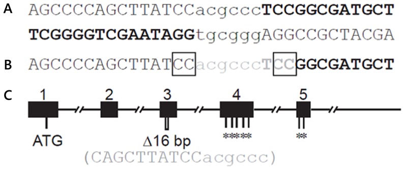
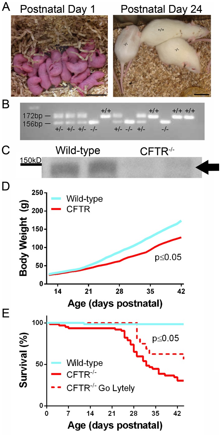
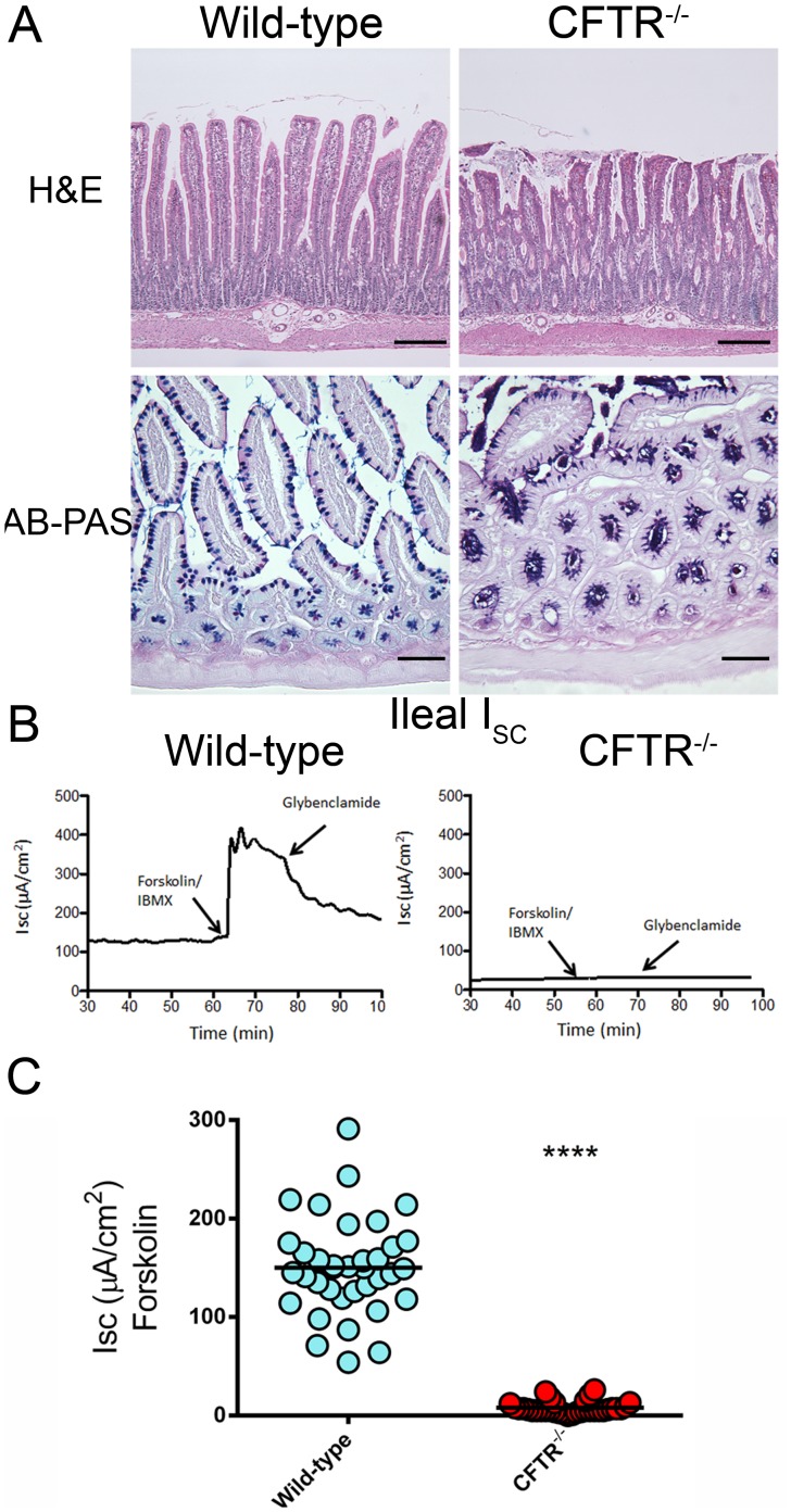
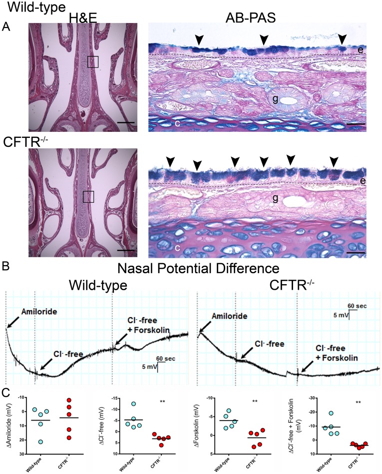
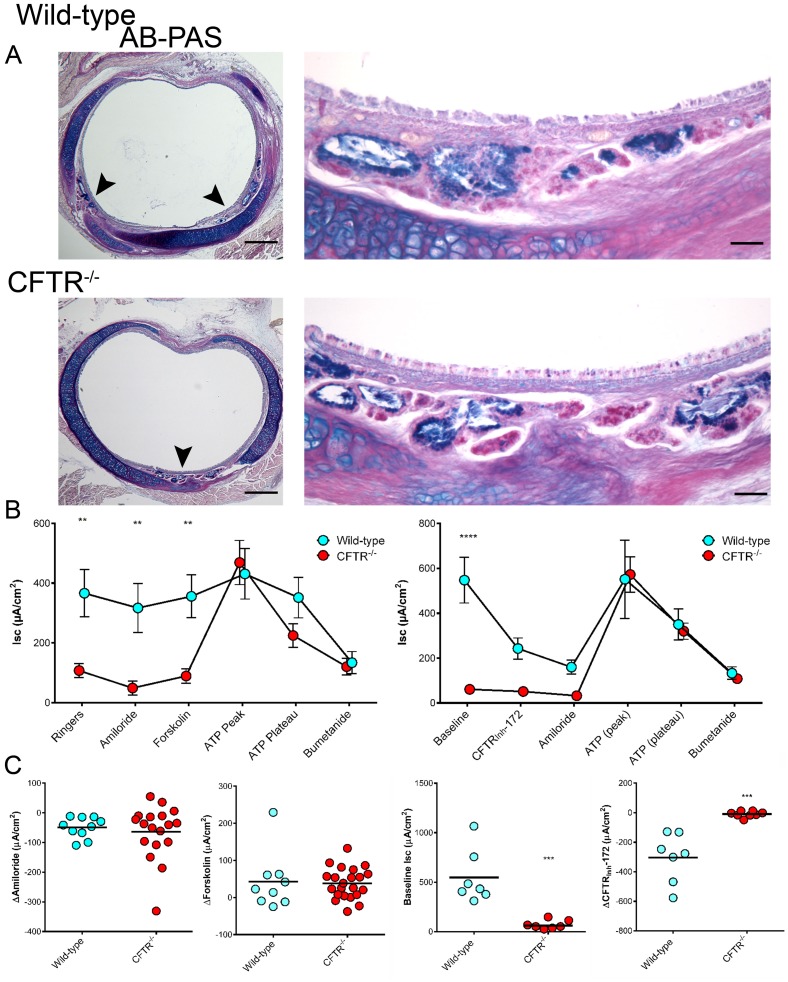
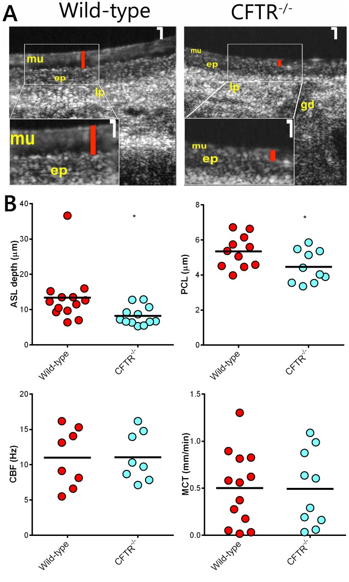
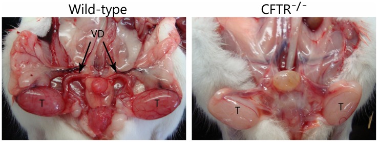
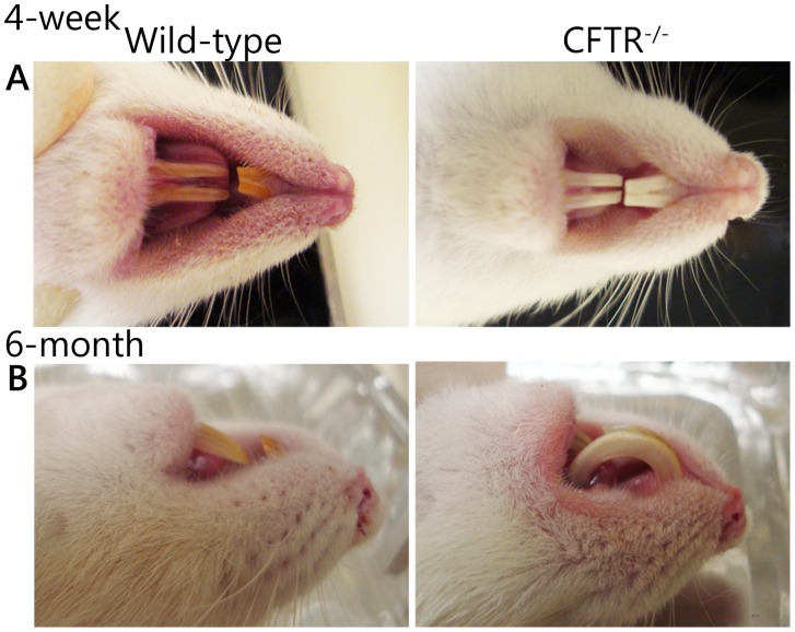
Animal models for cystic fibrosis (CF) have contributed significantly to our understanding of disease pathogenesis. Here we describe development and characterization of the first cystic fibrosis rat, in which the cystic fibrosis transmembrane conductance regulator gene (CFTR) was knocked out using a pair of zinc finger endonucleases (ZFN). The disrupted Cftr gene carries a 16 base pair deletion in exon 3, resulting in loss of CFTR protein expression. Breeding of heterozygous (CFTR+/-) rats resulted in Mendelian distribution of wild-type, heterozygous, and homozygous (CFTR-/-) pups. Nasal potential difference and transepithelial short circuit current measurements established a robust CF bioelectric phenotype, similar in many respects to that seen in CF patients. Young CFTR-/- rats exhibited histological abnormalities in the ileum and increased intracellular mucus in the proximal nasal septa. By six weeks of age, CFTR-/- males lacked the vas deferens bilaterally. Airway surface liquid and periciliary liquid depth were reduced, and submucosal gland size was abnormal in CFTR-/- animals. Use of ZFN based gene disruption successfully generated a CF animal model that recapitulates many aspects of human disease, and may be useful for modeling other CF genotypes, including CFTR processing defects, premature truncation alleles, and channel gating abnormalities.


Conflict of interest statement

Competing Interests: The authors have read the journal’s policy and have the following conflicts: XC, JW, LR, AC, DJ and KG are full-time employees of SAGE Labs, Inc., which sells genetically engineered rats and provides custom model creation services. KKC, EJS, SMR, and GT have filed a patent application related to use of µOCT technology in airways. The application serial number is PCT/US12/52553, entitled “Method for functional investigation of respiratory airways and other ciliated tissues using µOCT,” filed August 27, 2012. No financial benefit has yet been derived from this application by any author. SMR receives research support from the NIH, the Cystic Fibrosis Foundation (CFF), the American Lung Association, Synedgen Inc., and Forest Research Institute for basic and translational research. SMR also receives support from CFF Therapeutics (CFFT), Vertex Pharmaceuticals, PTC Therapeutics, N30 Therapeutics, and Novartis to conduct clinical trials related to the treatment and control of cystic fibrosis. SMR receives unrestricted research support for consulting services to Novartis, Kala Pharmaceuticals, Celtaxys Inc., and Bayer Healthcare. SMR receives no personal income from any of the sponsors noted above. GT receives research support from iLumen, Ninepoint Medical, Merck Research Laboratories, Canon, Samsung, and Air Liquide Medical Gases. GT. receives personal income from Ninepoint Medical, Terumo Corporation, and MIT through patent licensing arrangements. GT also receives consulting income from Samsung Advanced Institute of Technology and Ninepoint Medical. EJS receives research support from NIH, CFF, and CFFT. EJS serves as a scientific consultant to Genzyme, Pfizer, and Gilead. He receives consulting income from Gilead. KLT, SEB, JSH, LPT, JAF, MD, JMC, DMB, and MVF have no conflicts of interest to disclose. These disclosures do not alter the authors’ adherence to all PLOS ONE policies with regard to sharing data and materials.

Figures

Figure 1
Figure 1. Targeting in exon 3 of Cftr.
(A) ZFN recognition site sequence. The two ZFN binding sites are in bold uppercase. Cleavage site is in lower case. (B) Nine base pair deletion recovered in multiple founders. The deleted sequence is shown in gray. Microhomology that may have favored this deletion is marked in boxes. (C)Schematic of the gene structure of the first 5 exons of rat Cftr. Exons are shown by filled rectangles with exon number above. ATG, position of the translational start codon; Δ16 bp marks the position of 16 bp deletion, with nucleotide sequence below). *indicates premature stop mutations introduced by the 16 bp deletion in exon 3.
Figure 2
Figure 2. Generation of CFTR−/− rats.
(A) Animals at days 1 and 24 postnatal. (B) Results of PCR genotyping from a representative litter. (C) Western blot indicating expression of CFTR in wild-type rats and absence of CFTR protein from lungs of CFTR−/− animals. Arrow - CFTR. (D) Body weight values (mean ± SD) from wild-type and CFTR−/− rats from 12 to 44 days postnatal. (E) Survival curve for CFTR−/− rats from postnatal day 1 to 44 (p<0.05 for all groups, n = 12–67 animals/group).
Figure 3
Figure 3. Histology and short-circuit current measurements from small intestines of wild-type and CFTR−/− rats.
H&E (bar = 200 µm) and AB-PAS (bar = 100 µm) stained sections of the small intestines from wild-type and CFTR−/− rats. (n = 3–5 animals/group) (B) ISC tracings from wild-type and CFTR−/− rat ileum. (C) Summary of forskolin stimulated current measurements from ileal sections. (n = 5 animals/group) ****p≤0.0001.
Figure 4
Figure 4. Proximal nasal histology and nasal potential difference measurements.
(A) Low power magnification (4×) H&E stained sections from the proximal nasal passages bar = 500 µm. 20× images of ABPAS stained nasal septa from boxed areas bar = 25 µm. Arrowheads, cells swollen with intracellular mucus; e, respiratory epithelium; g, submucosal gland; dashed line (–), basement membrane (n = 4 animals/group) (B) NPD tracings from wild-type and CFTR−/− rats. (C) Summary data from NPD measurements for Δamiloride, ΔCl-free Ringers, Δforksolin, and ΔCl−/−free Ringers+forskolin. (n = 5 animals/group) **p≤0.01.
Figure 5
Figure 5. Tracheal histology and short circuit current measurements.
(A) AB-PAS stained tracheal sections from 6 week old rats. Submucosal glandular tissue indicated by arrowheads. Low magnification bar = 500 µm, high magnification bar = 50 µm (B & C) Summary data from Ussing chamber short circuit current measurements. Panels on right depict a modified protocol designed to specifically detect baseline (constitutive) CFTR function. (n = 3–6 animals/group) **p≤0.01, ***p≤0.001, ****p≤0.0001.
Figure 6
Figure 6. Functional anatomy of rat trachea.
(A) Representative time-averaged µOCT images of wild-type and CFTR−/− tracheas. Higher magnification insets (bottom left corner) show differences in ASL height between wild-type and CFTR−/− animals. White magnification bar = 10 µm. Red bars indicate ASL height. Mucus layer (mu), epithelium (ep), lamina propria (lp), and gland duct (gd) are also visualized. (B) Summary data for airway surface liquid depth (ASL), periciliary liquid depth (PCL), ciliary beat frequency (CBF), and mucociliary transport (MCT). (n = 8–13 animals/group) *p≤0.05.
Figure 7
Figure 7. Male CFTR−/− rats have bilateral absence of the vas deferens by 6 weeks of age.
Wild-type males (left) have an intact reproductive tract. CFTR−/− males (right) develop other reproductive organs, but exhibit absent vas at 6 weeks. T, testis; VD, vas deferens. (n = 3 animals/group).
Figure 8
Figure 8. CF rats have abnormal dentition.
(A) Wild-type rats (left) have yellowish-brown enamel while CFTR−/− rats (right) exhibit bright white incisors. (B) Incisors from CF rats exhibit uncontrolled growth and penetrate the hard palate. (n = 3 animals/group).

References

    1. Grosse SD, Boyle CA, Botkin JR, Comeau AM, Kharrazi M, et al. (2004) Newborn screening for cystic fibrosis: evaluation of benefits and risks and recommendations for state newborn screening programs. MMWR Recomm Rep 53: 1–36. - PubMed
    1. Derichs N (2013) Targeting a genetic defect: cystic fibrosis transmembrane conductance regulator modulators in cystic fibrosis. Eur Respir Rev 22: 58–65. - PMC - PubMed
    1. Riordan JR, Rommens JM, Kerem B, Alon N, Rozmahel R, et al. (1989) Identification of the cystic fibrosis gene: cloning and characterization of complementary DNA. Science 245: 1066–1073. - PubMed
    1. Guilbault C, Saeed Z, Downey GP, Radzioch D (2007) Cystic fibrosis mouse models. Am J Respir Cell Mol Biol 36: 1–7. - PubMed
    1. Carvalho-Oliveira I, Scholte BJ, Penque D (2007) What have we learned from mouse models for cystic fibrosis? Expert Rev Mol Diagn 7: 407–417. - PubMed

Publication types

MeSH terms

Substances