. 2014 Mar 7;9(3):e91089.
doi: 10.1371/journal.pone.0091089. eCollection 2014.

Anion exchange HPLC isolation of high-density lipoprotein (HDL) and on-line estimation of proinflammatory HDL

Affiliations

Anion exchange HPLC isolation of high-density lipoprotein (HDL) and on-line estimation of proinflammatory HDL

Xiang Ji et al. PLoS One. .

Abstract

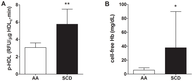
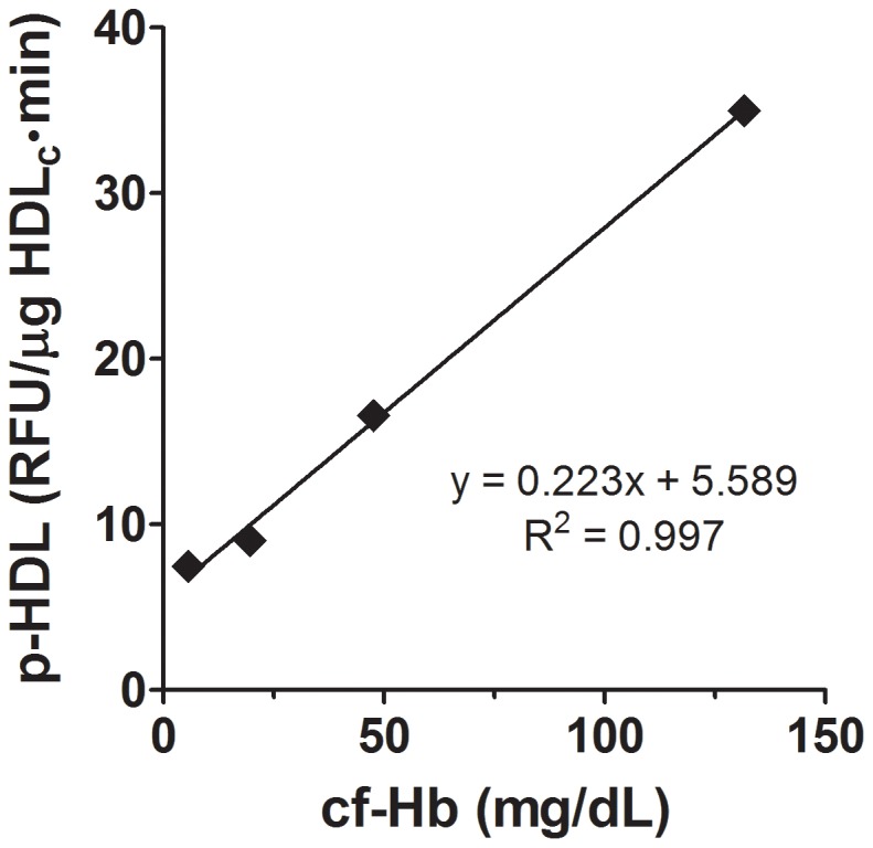
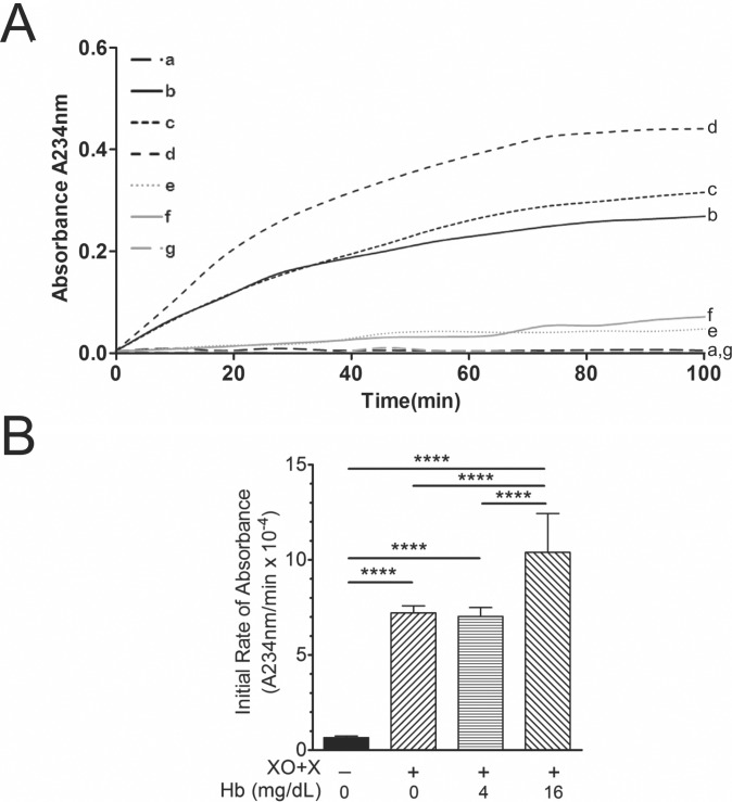
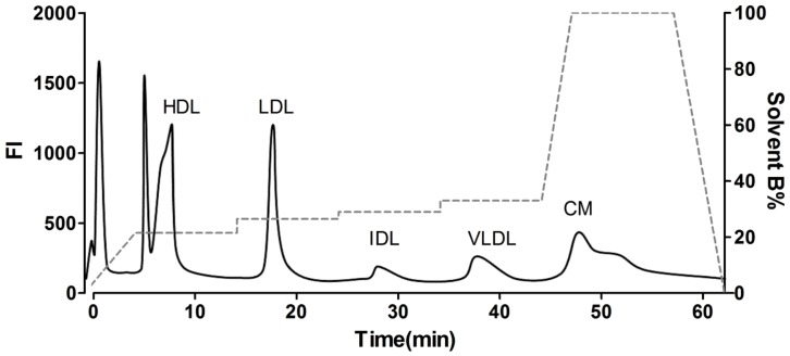
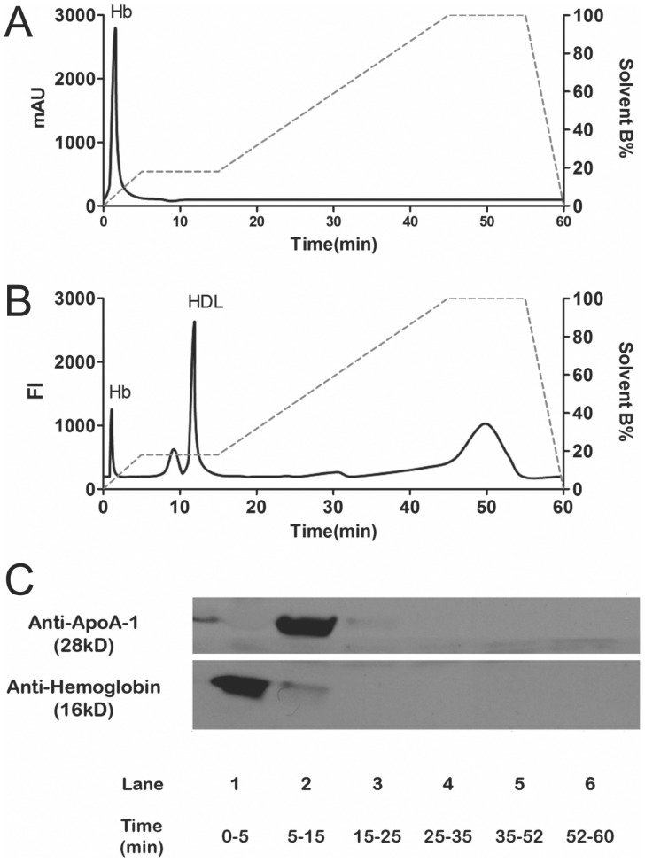
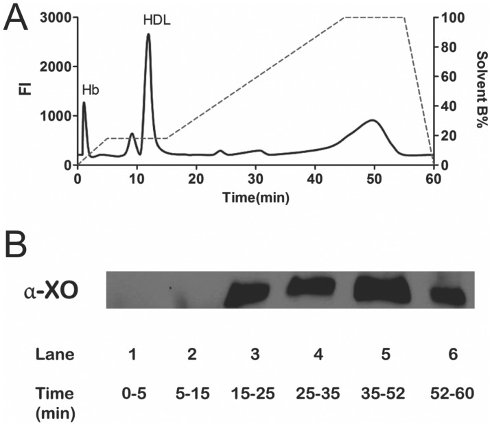
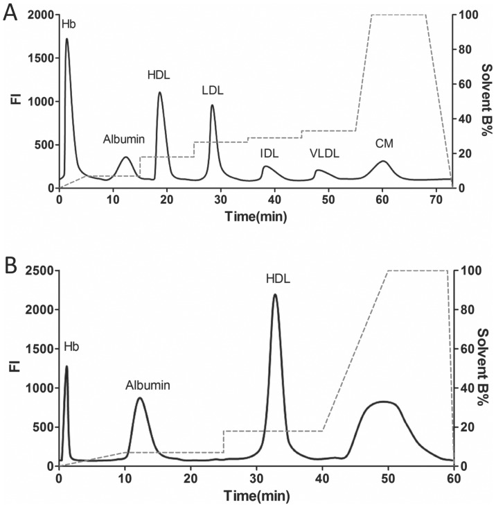
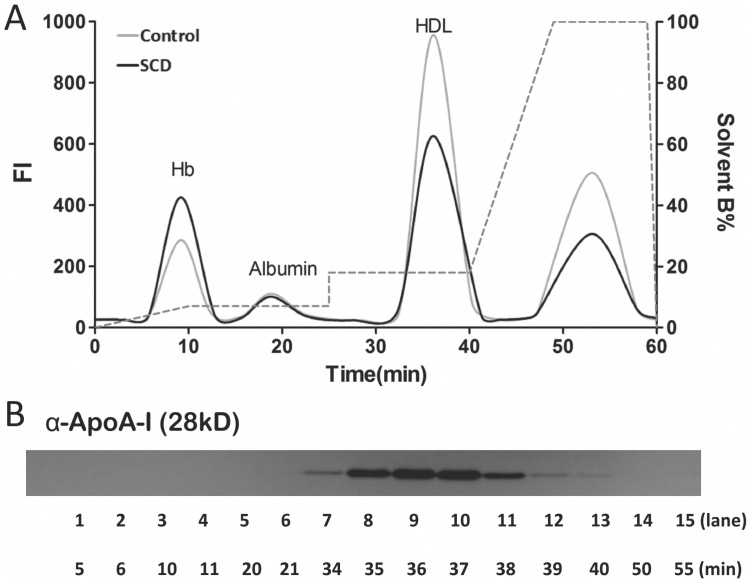
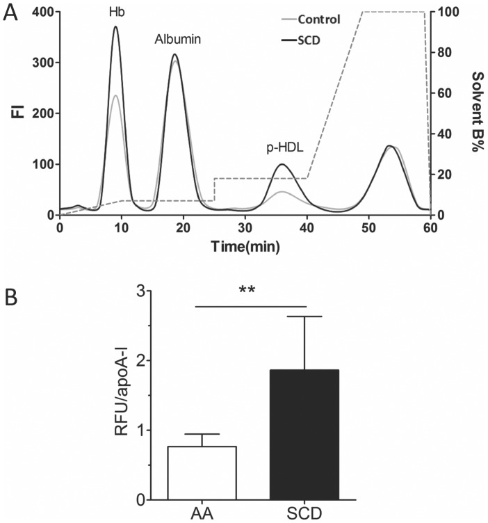
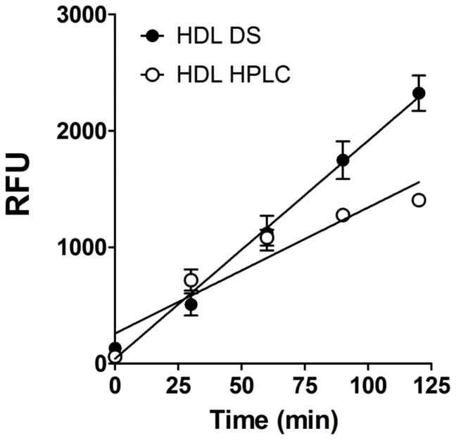
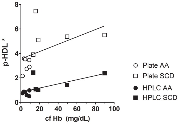
Proinflammatory high-density lipoprotein (p-HDL) is a biomarker of cardiovascular disease. Sickle cell disease (SCD) is characterized by chronic states of oxidative stress that many consider to play a role in forming p-HDL. To measure p-HDL, apolipoprotein (apo) B containing lipoproteins are precipitated. Supernatant HDL is incubated with an oxidant/LDL or an oxidant alone and rates of HDL oxidation monitored with dichlorofluorescein (DCFH). Although apoB precipitation is convenient for isolating HDL, the resulting supernatant matrix likely influences HDL oxidation. To determine effects of supernatants on p-HDL measurements we purified HDL from plasma from SCD subjects by anion exchange (AE) chromatography, determined its rate of oxidation relative to supernatant HDL. SCD decreased total cholesterol but not triglycerides or HDL and increased cell-free (cf) hemoglobin (Hb) and xanthine oxidase (XO). HDL isolated by AE-HPLC had lower p-HDL levels than HDL in supernatants after apoB precipitation. XO+xanthine (X) and cf Hb accelerated purified HDL oxidation. Although the plate and AE-HPLC assays both showed p-HDL directly correlated with cf-Hb in SCD plasma, the plate assay yielded p-HDL data that was influenced more by cf-Hb than AE-HPLC generated p-HDL data. The AE-HPLC p-HDL assay reduces the influence of the supernatants and shows that SCD increases p-HDL.


Conflict of interest statement

Competing Interests: The authors have declared that no competing interests exist.

Figures

Figure 1
Figure 1. Effects of SCD on p-HDL and cf Hb.
(A) Proinflammatory HDL (p-HDL) in SCD patients was increased compared with controls (** = p<0.01, n = 6). (B) Cell-free (cf) Hb was increased in SCD patients compared with controls (* = p<0.05, n = 6).
Figure 2
Figure 2. Effects of cf Hb on p-HDL.
P-HDL levels were determined by the plate assay. Pooled mouse plasma was analyzed for cf Hb (5.6 mg/dL) and p-HDL (7.46 RFU/min). Increasing concentrations of human Hb were added to the mouse plasma, apoB lipoproteins precipitated and supernatant p-HDL levels determined immediately after apo B precipitation. Plotted data were mean of duplicates.
Figure 3
Figure 3. Effects of XO, X, cf Hb alone and in combination on HDL conjugated diene formation.
(A) XO, X, cf Hb alone and in combination were added to purified human HDL and initial rates of absorbance (A234 nm) recorded over time. Absorbance at 234 nm was recorded and initial rates of absorbance calculated per test group. (A) Line graphs showing changes in A234 nm with respect to time for the following test groups: a) HDL alone; b) HDL+XO+X; c) HDL+Hb (4 mg/dL) +XO+X; d) HDL+Hb (16 mg/dL) +XO+X; e) HDL+X; f) HDL+XO; and, g) XO+X. (B) The bar chart showed that XO/X increases A234 nm at faster rates than HDL alone and that adding cf Hb increased initial rates of HDL oxidation greater than XO/X alone when cf Hb equals 16 mg/dL but, not 4 mg/dL. (**** = p<0.001, n = 5–12).
Figure 4
Figure 4. AE-HPLC Protocol for Separating Lipoproteins.
DiI (10 µg/mL, final concentration) was added to plasma and lipoproteins separated by AE-HPLC using the protocol published by Hirowatari et al.20 This DiI chromatogram shows that all 5 classes of lipoproteins are separated as reported.20 However, the chromatogram also shows that DiI associates with two other components, at the very front of the chromatogram and in a peak on the shoulder of the HDL peak.
Figure 5
Figure 5. AE-HPLC separation of cf Hb and HDL from plasma.
AE-HPLC of Hb alone revealed Hb elutes at 0–5 min. (A) Cf Hb (100 mg/dL) 30 µl was injected without DiI, which was measured by UV absorbance (A230 nm). (B) Plasma treated with DiI (10 µg/mL) and then 30 µL was analyzed by fluorescent AE-HPLC (Ex 530 nm/Em 577 nm). (C) Plasma (30 µL) was injected, fractions collected, pooled, concentrated and examined by immunoblot analysis for apoA-I and Hb.
Figure 6
Figure 6. Separation of Xanthine Oxidase (XO) from HDL.
DiI-treated plasma was injected (30 µL) into the AE-HPLC. Fractions were collected, concentrated and examined by immunoblot for XO. The chromatogram in A and immunoblot in B showed that the modified AE-HPLC protocol eluted HDL prior to XO.
Figure 7
Figure 7. Standardized Lipoprotein and HDL Elution Profiles.
(A) DiI chromatogram for AE-HPLC separation of Hb, Albumin and the 5 lipoprotein (HDL, LDL, IDL, VLDL, CM) fractions. (B) DiI chromatogram for AE-HPLC separation of Hb, Albumin and HDL.
Figure 8
Figure 8. Effects of post-column reactor (PCR) on AE-HPLC separation of Hb, albumin and HDL.
(A) DiI chromatogram showing that HDL can be separated from Hb, albumin and other lipoproteins albeit with broadened peaks. (B) Anti-apoA-I immunoblot of pooled fractions showing that apoA-I (i.e., HDL) eluted between 34 and 39 minutes in the modified protocol.
Figure 9
Figure 9. AE-HPLC-PCR Quantification of p-HDL in Control (AA) and SCD Plasma.
(A) DCF chromatogram showing relative levels of DCF fluorescence (index of oxidizability) for p-HDL in control and SCD plasma. At 30–44 minutes, DCF fluorescence intensity in separated HDL in SCD plasma (black line) is greater than in control plasma (gray line). (B) Dividing DCF fluorescence intensity under these peaks by the subject’s plasma apoA-I concentration (µg/mL) yields relative fluorescence units (RFU) per apoA-I (µg/mL). P-HDL are increased in SCD subjects compared to control subjects (** = p<0.01, n = 6).
Figure 10
Figure 10. Effects of different methods for isolating HDL on rates of HDL oxidation determined by the plate assay.
HDL was isolated from plasma by dextran sulfate-MgCl2 precipitation of apo B lipoproteins (black-filled circles, DS) or by AE-HPLC (white open circles). Rates of DCF oxidation in HDL isolated by DS and HPLC were determined by the plate assay (equal quantities of HDL cholesterol). Data were presented as mean ± SD of assays performed in triplicate. (y = ax+b; HDL DS: y = 18.728x+42.794, R2 = 0.9934; HDL HPLC: y = 10.817x+259.46 R2 = 0.9035).
Figure 11
Figure 11. P-HDL plotted as a function of cf Hb: Plate vs. HPLC Assay.
For plate assay, control AA p-HDL data (* RFU/µg HDLc⋅min) were plotted as open circles and SCD p-HDL data were plotted as open squares. For HPLC assay, controls (AA, meaning homozygous for hemoglobin AA) p-HDL data (* RFU/apoA-I (µg/mL)) were plotted as closed circles and SCD p-HDL data were plotted as closed squares. (y = ax+b; Plate assay: y = 0.03004x+3.554; HPLC assay: y = 0.01688x+0.84140). These data showed that p-HDL isolated by apo B precipitation was more sensitive to the effects of cf Hb and was more variable than p-HDL determined by the AE-HPLC-PCR assay.

Similar articles

Cited by

References

    1. Assmann G, von Eckardstein A, Funke H (1993) High density lipoproteins, reverse transport of cholesterol, and coronary artery disease. Insights from mutations. Circulation 87: III28–34. - PubMed
    1. Navab M, Fogelman AM, Berliner JA, Territo MC, Demer LL, et al. (1995) Pathogenesis of atherosclerosis. Am J Cardiol 76: 18C–23C. - PubMed
    1. Gharavi NM, Gargalovic PS, Chang I, Araujo JA, Clark MJ, et al. (2007) High-density lipoprotein modulates oxidized phospholipid signaling in human endothelial cells from proinflammatory to anti-inflammatory. Arterioscler Thromb Vasc Biol 27: 1346–1353. - PubMed
    1. deGoma EM, deGoma RL, Rader DJ (2008) Beyond high-density lipoprotein cholesterol levels evaluating high-density lipoprotein function as influenced by novel therapeutic approaches. J Am Coll Cardiol 51: 2199–2211. - PMC - PubMed
    1. Khera AV, Cuchel M, de la Llera-Moya M, Rodrigues A, Burke MF, et al. (2011) Cholesterol efflux capacity, high-density lipoprotein function, and atherosclerosis. N Engl J Med 364: 127–135. - PMC - PubMed

Publication types

MeSH terms

LinkOut - more resources