Review
. 2014 Mar;7(3):331-41.
doi: 10.1242/dmm.014589.

Glycoprotein folding and quality-control mechanisms in protein-folding diseases

Affiliations
Review

Glycoprotein folding and quality-control mechanisms in protein-folding diseases

Sean P Ferris et al. Dis Model Mech. 2014 Mar.

Abstract

Biosynthesis of proteins--from translation to folding to export--encompasses a complex set of events that are exquisitely regulated and scrutinized to ensure the functional quality of the end products. Cells have evolved to capitalize on multiple post-translational modifications in addition to primary structure to indicate the folding status of nascent polypeptides to the chaperones and other proteins that assist in their folding and export. These modifications can also, in the case of irreversibly misfolded candidates, signal the need for dislocation and degradation. The current Review focuses on the glycoprotein quality-control (GQC) system that utilizes protein N-glycosylation and N-glycan trimming to direct nascent glycopolypeptides through the folding, export and dislocation pathways in the endoplasmic reticulum (ER). A diverse set of pathological conditions rooted in defective as well as over-vigilant ER quality-control systems have been identified, underlining its importance in human health and disease. We describe the GQC pathways and highlight disease and animal models that have been instrumental in clarifying our current understanding of these processes.

Keywords: ER export; ER quality control; ER-associated degradation; Glycoprotein folding; N-glycosylation.


Figures
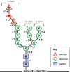
Fig. 1.
Fig. 1.
Structure of the N-linked core glycan. The triantennary tetradecaoligosaccharide is assembled on the ER membrane and is covalently linked to the Asn side chains in the context of the N-glycosylation sequon of newly translocated proteins. The 14-sugar form, starting from the Asn residue, contains two N-acetylglucosamine (GlcNAc, squares), nine mannose (circles) and three glucose (triangles) residues. The three branches – A, B and C – are illustrated. Glycosidic linkage types are indicated next to the connectors.
Fig. 2.
Fig. 2.
N-glycan processing in the CNX cycle. Glycoproteins first enter the CNX cycle after the two terminal glucose residues (red triangles) of the attached N-glycan are cleaved by glucosidases I and II (GS-I and GS-II; steps 1 and 2). The resulting monoglucosylated N-glycan binds to the lectin-like chaperones CNX and CRT. The substrate dissociates from CNX/CRT upon GS-II-mediated removal of the terminal glucose residue from the N-glycan (step 3). At this point, the glycoprotein substrate’s folding status is surveyed by the ‘folding sensor’ component of the CNX cycle, UGGT1, which specifically binds nearly-native folding forms (step 4) and reglucosylates them. Reglucosylated substrates bind to CNX/CRT once again and re-enter the CNX cycle (step 5). Substrates eventually exit the CNX cycle upon demannosylation (removal of mannose residues; green circles) of N-glycans (step 6). The mechanism for permanent exit from the cycle involves either termination of UGGT1 reglucosylation activity of demannosylated N-glycans, or active recognition of demannosylated forms by ER exit machinery or ERAD components.
Fig. 3.
Fig. 3.
Glycoprotein ERAD. Degradation of terminally misfolded glycoproteins through ERAD is probably initiated by cleavage of the terminal B-chain mannose (green circles) of Man9GlcNAc2 (and possibly Glc1Man9GlcNAc2) N-glycan forms by ER mannosidase I (ERManI, step 1). This results in the formation and recognition of this specific Man8GlcNAc2 form by ER-degradation-enhancing alpha-mannosidase-like 1 (EDEM1). Then, removal of the terminal C-chain mannose, either: (1) directly by EDEM3 or possibly Golgi mannosidase I (step 2), or (2) by highly concentrated ERMan I in the ERQC compartment (step 3), exposes an α1-6 linked mannose that is recognized by OS-9. OS-9 facilitates transport of the misfolded substrate to the core ERAD HRD1-SEL1L complex (step 4), and subsequent retrotranslocation to the cytoplasm for degradation by the proteasome (step 5).
Fig. 4.
Fig. 4.
Degradation of insoluble and soluble ER glycoproteins. Some misfolded glycoproteins form insoluble aggregates or ordered polymers in the ER, and UGGT1-mediated modification of the glucosylation status (monoglucosylated or unglucosylated) might play a role in limiting insolubility. The soluble-to-insoluble transition might occur via compartmentalization in the ERAC, and insoluble substrates might also be resolubilized. Soluble forms of both luminal and transmembrane glycoproteins tend to be degraded through ERAD, whereas insoluble forms tend to be degraded via autophagy, although the mechanism by which insoluble ER proteins get to the lysosome is not entirely clear. Luminal insoluble glycoproteins might be packaged into EDEM1-containing vesicles (EDEMosomes) and transported to the lysosome. Transmembrane insoluble glycoproteins could accumulate in the aggresome and then be degraded by the proteasome, or be targeted by an unknown mechanism to the lysosome for degradation.

References

    1. Aebi M., Bernasconi R., Clerc S., Molinari M. (2010). N-glycan structures: recognition and processing in the ER. Trends Biochem. Sci. 35, 74–82 - PubMed
    1. Appenzeller C., Andersson H., Kappeler F., Hauri H. P. (1999). The lectin ERGIC-53 is a cargo transport receptor for glycoproteins. Nat. Cell Biol. 1, 330–334 - PubMed
    1. Appenzeller-Herzog C., Roche A. C., Nufer O., Hauri H. P. (2004). pH-induced conversion of the transport lectin ERGIC-53 triggers glycoprotein release. J. Biol. Chem. 279, 12943–12950 - PubMed
    1. Appenzeller-Herzog C., Nyfeler B., Burkhard P., Santamaria I., Lopez-Otin C., Hauri H. P. (2005). Carbohydrate- and conformation-dependent cargo capture for ER-exit. Mol. Biol. Cell 16, 1258–1267 - PMC - PubMed
    1. Apweiler R., Hermjakob H., Sharon N. (1999). On the frequency of protein glycosylation, as deduced from analysis of the SWISS-PROT database. Biochim. Biophys. Acta 1473, 4–8 - PubMed

Publication types

LinkOut - more resources