. 2014 May;42(9):5505-17.
doi: 10.1093/nar/gku183. Epub 2014 Mar 7.

Myc-induced anchorage of the rDNA IGS region to nucleolar matrix modulates growth-stimulated changes in higher-order rDNA architecture

Affiliations

Myc-induced anchorage of the rDNA IGS region to nucleolar matrix modulates growth-stimulated changes in higher-order rDNA architecture

Chiou-Nan Shiue et al. Nucleic Acids Res. 2014 May.

Abstract

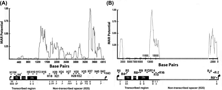
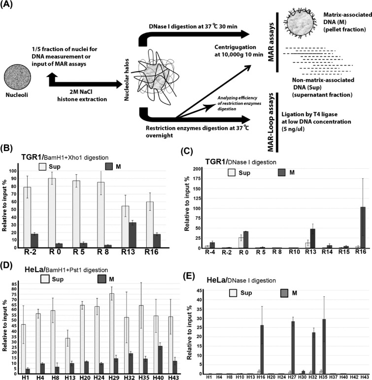
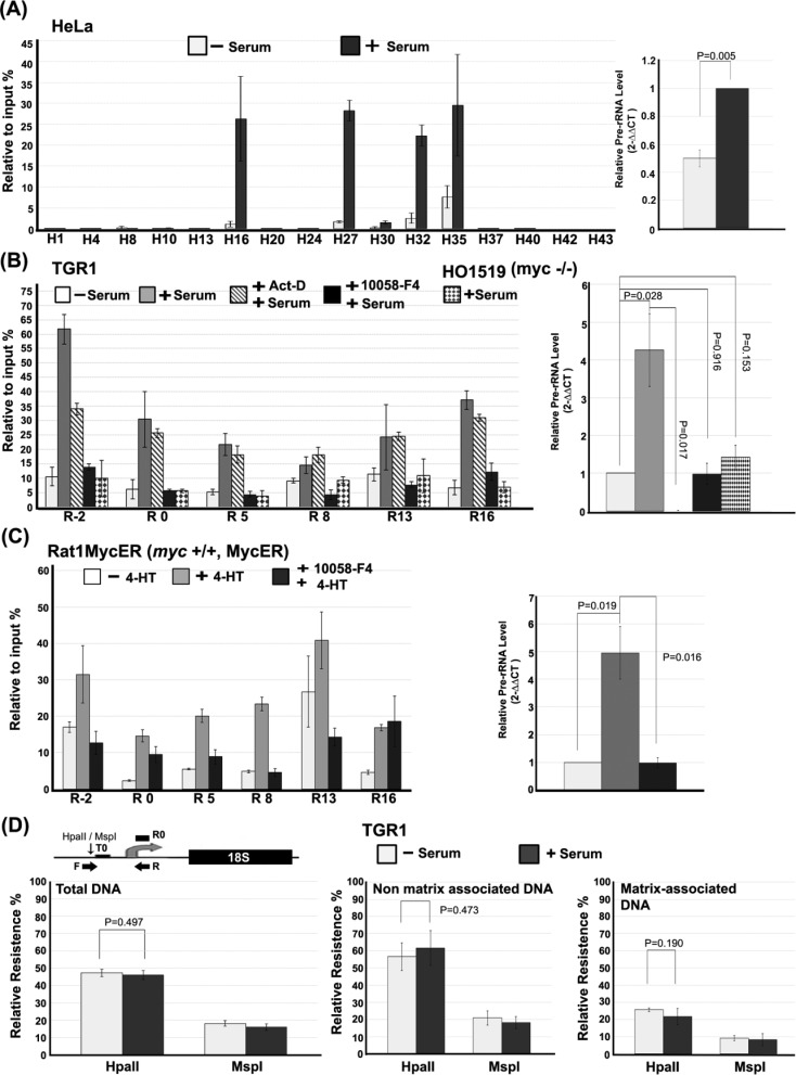
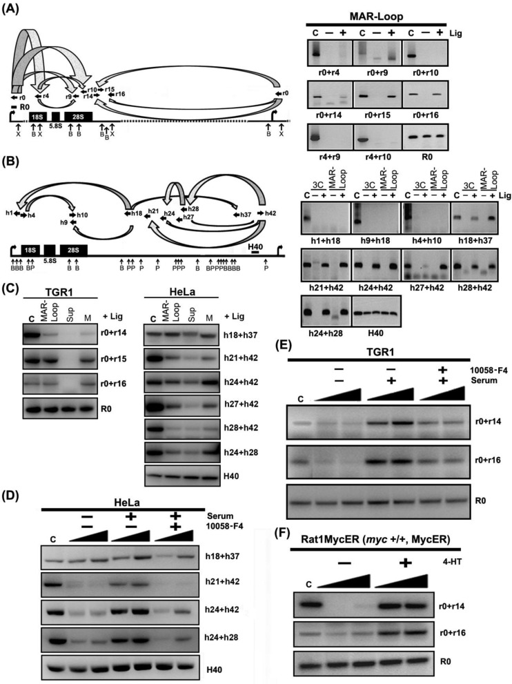
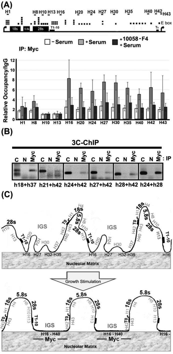
Chromatin domain organization and the compartmentalized distribution of chromosomal regions are essential for packaging of deoxyribonucleic acid (DNA) in the eukaryotic nucleus as well as regulated gene expression. Nucleoli are the most prominent morphological structures of cell nuclei and nucleolar organization is coupled to cell growth. It has been shown that nuclear scaffold/matrix attachment regions often define the base of looped chromosomal domains in vivo and that they are thereby critical for correct chromosome architecture and gene expression. Here, we show regulated organization of mammalian ribosomal ribonucleic acid genes into distinct chromatin loops by tethering to nucleolar matrix via the non-transcribed inter-genic spacer region of the ribosomal DNA (rDNA). The rDNA gene loop structures are induced specifically upon growth stimulation and are dependent on the activity of the c-Myc protein. Matrix-attached rDNA genes are hypomethylated at the promoter and are thus available for transcriptional activation. rDNA genes silenced by methylation are not recruited to the matrix. c-Myc, which has been shown to induce rDNA transcription directly, is physically associated with rDNA gene looping structures and the intergenic spacer sequence in growing cells. Such a role of Myc proteins in gene activation has not been reported previously.


Figures

Figure 1.
Figure 1.
The non-transcribed intergenic spacer (IGS) region of rRNA genes has an enhanced propensity for nuclear matrix attachment. The line graphs show MAR potential score in relation to nucleotide position for the rDNA repeat sequence from human (A) and rat (B). The lower panels in each figure part show schematic depictions of rRNA gene repeats. Vertical arrows below the rDNA repeat represent the locations of cleavage sites for BamHI (B) and PstI (P) in human (part A) and for BamHI (B) and XhoI (X) in rat (part B). The locations of amplified rDNA regions are shown by horizontal bars. The sequences of primer sets used for amplification of human and rat rDNA are shown in Supplementary Tables S1 and S2. The dotted line in rat rRNA gene represents sequences that are not available from GenBank. Human rDNA sequence, derived from GenBank number U13369, and (B) rat rDNA sequence, derived from accession numbers X04084, X03838, X61110, X00677, X16321, V01270 and X03695, were used. Matrix attachment potential was predicted by the MAR-Wiz software (57) as implemented at http://genomecluster.secs.oakland.edu using a window size of 1000 bp stepped at 100-bp intervals. Scores in excess of 0.6 are generally considered as strong indicators of nuclear matrix binding potential.
Figure 2.
Figure 2.
Preferential matrix attachment of rDNA genes via the intergenic spacer (IGS) region. (A) Diagram showing the procedure used for isolation of nucleolar halos and the preparation of nucleolar matrix and subsequent MAR and MAR-loop assays. (B) Preferential attachment of rDNA genes to nuclear matrix via IGS sequences in growing TGR-1 cells. The relative association of different regions of the rDNA repeat to nucleolar matrix (M) after digestion with BamH1 and Xho1 as well as the levels of released non-matrix associated regions (Sup) are shown. (C) Fine mapping of nucleolar-matrix-associated rDNA fragments after DNase I treatment of nucleolar matrix from growing TGR-1 cells. (D) Preferential attachment of rDNA genes to nuclear matrix via sequences throughout the IGS region in growing HeLA cells. The supernatant fraction and pellet fraction are separated after restriction enzymes (BamH1+Pst1) digestion. (E) Fine mapping of nucleolar-matrix-associated rDNA fragments after DNase I treatment of nucleolar matrix from growing HeLa cells. The values plotted in (B–E) are the means and standard deviation of results from three independent experiments.
Figure 3.
Figure 3.
Growth-dependent and c-Myc-dependent attachment of rDNA to the nucleolar matrix. (A) Matrix attachment of the rDNA IGS is induced upon growth stimulation of HeLa cells. The left panel represents quantitative real-time PCR showing the relative levels of matrix-attached rDNA throughout the rDNA repeat after digestion with DNase I for starved HeLa cells before (−S) or after (+S) re-feeding with serum-containing medium. The right panel shows the corresponding changes in the level of pre-rRNA measured by quantitative real-time PCR. (B) Growth-induced attachment of the rDNA IGS to nuclear matrix requires c-Myc in rat fibroblasts. Levels of matrix attachment after restriction digestion (see Figure 2B) are shown for starved TGR-1 cells re-fed with serum in the absence or presence of actinomycin D (0.1 μg/ml) or the Myc inhibitor, 10058-F4 (80 μM). Growing HO1519 (myc−/−) cells were treated and analyzed in parallel. Pre-rRNA levels corresponding to the different cells and treatments are shown (right panel). (C) Activation of Myc in serum-starved cells is sufficient to induce rDNA IGS matrix attachment. The relative levels of matrix-attached rDNA after restriction digestion (see Figure 2B) at the indicated rDNA regions in Rat1MycER cells, which express a Myc-ER fusion protein, before (−4-HT) and after (+4-HT) activation of Myc-ER by addition of 4-hydroxytamoxifen in the absence or presence of c-Myc inhibitor 10058-F4. Pre-rRNA levels corresponding to the different treatments are shown (right panel). The cutting efficiencies of restriction enzymes on samples in (B) and (C) are shown in Supplementary Figure S2D and E. (D) rRNA genes that associate with nucleolar matrix are hypomethylated in the promoter region. A diagram of the rat rDNA promoter region shows the primer set (forward primer, F; reverse primer, R), described in the Materials and Methods section, used to detect the methylation status of the CpG residue at −145 bp from the transcription start site, while the primer set R0 is for normalization between samples (upper panel; see the Materials and Methods section). The quantitative real-time PCR signal for genomic DNA from serum-starved (−Serum) and growing (+ Serum) TGR-1 cells after cleavage with HpaII or MspI, prior to (left panel) or after separation of non-matrix-associated DNA (middle panel) and matrix-associated DNA (right panel). The values in (A–D) are the means and standard deviations of results from three independent experiments.
Figure 4.
Figure 4.
rDNA IGS matrix attachment can account for growth- and Myc-dependent changes in higher-order rDNA structure. (A) Distant regions within IGS are bound to matrix in close proximity to each other. The left panel shows the combinations of primer sets used and their locations and orientations along rat rDNA repeat (sequences of primer sets are shown in Supplementary Table S4). The right panels show results for different primer pairs from an MAR-loop assay of growing TGR-1 cells, in which DNA fragments, which are held in close proximity to each other after cleavage with XhoI (X) and BamHI (B) by matrix attachment, can be ligated (+ Lig) to create novel DNA fragments. The panels also show the migration of positive control (C) fragments, which indicate the expected size of potential ligation products, and that no novel DNA fragments are detected in the absence of added ligase (− Lig). The last panel (R0) is a loading control (see Figure 1B). (B) The MAR-loop assay and 3C assay identify the involvement of an equivalent set of rDNA IGS regions in the formation of rDNA gene loop structures in growing HeLa cells. Annotations are the same as for part (A) (sequences of primer pairs are shown in Supplementary Table S6) except that the H40 region is amplified in all samples as a loading control. (C) Positive proximity results from the MAR-loop assay are predominately associated with the matrix fraction when the assay is performed on isolated matrix-associated (M) and matrix-non-associated (Sup) fractions. Other annotations are shown as (A) and (B). Growth stimulation of matrix-associated gene loop structures is dependent on the activity of c-Myc in (D) HeLa cells and (E) TGR-1 cells. MAR-ligation assay results for starved HeLa and TGR-1 cells before (−serum) or after (+serum) addition of medium containing serum in the absence or presence of c-Myc inhibitor, 10058-F4. Other annotations are as for part (A) and part (B). The filled ramps indicate that PCR amplifications were performed at increasing substrate concentrations, since product formation is easily saturated at higher product concentrations. (F) Myc-ER activation is sufficient to induce matrix-associated gene looping in cells lacking endogenous c-Myc. MAR-ligation assay results before (−4-HT) or after (+4-HT) treatment of Rat1MycER cells. The cutting efficiencies of restriction enzymes on all the indicated samples are shown in Supplementary Figure S2C–E.
Figure 5.
Figure 5.
c-Myc associates with the rDNA IGS region in a growth-dependent fashion and is physically associated with the gene looping structures that are found in growing cells. (A) Quantitative real-time PCR ChIP assays to detect association of c-Myc with rDNA in starved HeLa cells before (−serum) and after addition of serum (+serum) or serum and 10058-F4 (+serum, +10058-F4). The upper panel shows a human rDNA repeat marked with locations of E boxes (canonical and non-canonical) and primer pairs for qPCR. Binding values are expressed relative to levels detected after parallel ChIP reactions with IgG from non-immunized rabbit. Error bars show standard deviation about the mean (n ≥ 4). (B) c-Myc is physically associated with rDNA-IGS-mediated gene looping structures in growing HeLa cells. The re-ligated rDNA chromatin, immunoprecipitated by a specific antibody (Myc) or agarose beads as a negative control (N), is amplified by using primer pairs throughout the rDNA IGS region (sequences of primer sets are shown in Supplementary Table S6). The expected migration of the ligation product is shown by the positive control lane (C). (C) A schematic diagram describing the main conclusions of the work. Hypomethylated (potentially active) rDNA genes associate with nucleolar matrix via the IGS in a growth- and Myc-dependent manner. Matrix association appears to be the cause of the growth-related gene looping structures that characterize the rDNA in growing cells. Myc is associated with the growth-stimulated gene looping structures via interaction with the rDNA IGS.

Similar articles

Cited by

References

    1. Felsenfeld G. Chromatin unfolds. Cell. 1996;86:13–19. - PubMed
    1. Brown K.E. Chromatin folding and gene expression: new tools to reveal the spatial organization of genes. Chromosome Res. 2003;11:423–433. - PubMed
    1. Mehta I.S., Figgitt M., Clements C.S., Kill I.R., Bridger J.M. Alterations to nuclear architecture and genome behavior in senescent cells. Ann. N.Y. Acad. Sci. 2007;1100:250–263. - PubMed
    1. Prieto J.L., McStay B. Nucleolar biogenesis: the first small steps. Biochem. Soc. Trans. 2005;33:1441–1443. - PubMed
    1. Raska I., Shaw P.J., Cmarko D. New insights into nucleolar architecture and activity. Int. Rev. Cytol. 2006;255:177–235. - PubMed

Publication types

Substances