. 2014 Apr 1;192(7):3080-90.
doi: 10.4049/jimmunol.1301053. Epub 2014 Mar 7.

In vivo detection of peripherin-specific autoreactive B cells during type 1 diabetes pathogenesis

Affiliations

In vivo detection of peripherin-specific autoreactive B cells during type 1 diabetes pathogenesis

Nahir Garabatos et al. J Immunol. .

Abstract

Autoreactive B cells are essential for the pathogenesis of type 1 diabetes. The genesis and dynamics of autoreactive B cells remain unknown. In this study, we analyzed the immune response in the NOD mouse model to the neuronal protein peripherin (PRPH), a target Ag of islet-infiltrating B cells. PRPH autoreactive B cells recognized a single linear epitope of this protein, in contrast to the multiple epitope recognition commonly observed during autoreactive B cell responses. Autoantibodies to this epitope were also detected in the disease-resistant NOR and C57BL/6 strains. To specifically detect the accumulation of these B cells, we developed a novel approach, octameric peptide display, to follow the dynamics and localization of anti-PRPH B cells during disease progression. Before extended insulitis was established, anti-PRPH B cells preferentially accumulated in the peritoneum. Anti-PRPH B cells were likewise detected in C57BL/6 mice, albeit at lower frequencies. As disease unfolded in NOD mice, anti-PRPH B cells invaded the islets and increased in number at the peritoneum of diabetic but not prediabetic mice. Isotype-switched B cells were only detected in the peritoneum. Anti-PRPH B cells represent a heterogeneous population composed of both B1 and B2 subsets. In the spleen, anti-PRPH B cell were predominantly in the follicular subset. Therefore, anti-PRPH B cells represent a heterogeneous population that is generated early in life but proliferates as diabetes is established. These findings on the temporal and spatial progression of autoreactive B cells should be relevant for our understanding of B cell function in diabetes pathogenesis.


Figures
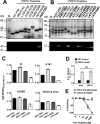
Figure 1
Figure 1. Identification of a C-terminal epitope recognized by islet-infiltrating anti-PRPH B cells
(A) SDS PAGE analysis of purified overlapping PRPH peptides (top) and WB analysis of the same fragments using mAb I6 as primary antibody (bottom). The recognized peptide is highlighted in bold (see also Figure S1). (B) Fine-mapping of the epitope recognized by mAb I6. SDS PAGE analysis (top) and WB analysis of the same fragments using I6 as primary antibody (bottom). Crude E. coli protein extracts from induced cultures were analyzed. The recognized peptides are highlighted in bold (see also Table 1). (C) Differential recognition of peptides PRPH429-507, PRPH467-507, PRPH473-495, and PRPH475-491 by representative B cell hybridomas and sera collected from 19-wk-old NOD mice. Peptides were coated to ELISA plates, incubated with culture supernatants from the hybridoma cultures or mouse sera, and specific Abs detected using anti-mouse IgG. Values of the GST blank control were subtracted during analysis. Sera dilution: 1/300. (D) mAb I6 recognizes a linear epitope. ELISA analysis of coated PRPH429-507 and GST as a control, both treated with urea or left untreated. Bound mAb was detected as in (C). Data (mean ± SD) are representative of 3 independent experiments. The P value was determined by Mann Whitney t test, ns (no significant) P > 0.05. (E) mAb I6 recognizes a synthetic version of PRPH473-495. An ELISA competition assay is shown, using plate-bound GST-PRPH473-495 and free, synthetic peptide PRPH473-495 for competition. mAb I6 was preincubated using the indicated concentrations of free peptide overnight and next added to coated plates. Bound mAb was detected as in (C). The irrelevant synthetic PRPH435-449 peptide was used as a control in this assay. Data (mean ± SD) are representative of 3 independent experiments.
Figure 2
Figure 2. Anti-PRPH humoral response in diabetes-prone and diabetes-resistant strains
ELISA detection of anti-PRPH IgG in sera harvested from NOD, NOR, [NODxNOR] F1 hybrids, and C57BL/6 mice. For detection, PRPH429-507 was coated to plates. RU (relative units) values were normalized to an internal anti-hexa-histidine Ab control run in each experiment to allow cross-plate comparison of data. Five animals per group of sex and age were used. Sera dilution: 1/300. Data are the mean ± SD of 3-5 mice in each group. The P value was determined by 1-way ANOVA test for each female and male group. ***P < 0.001, ** P < 0.01 *P < 0.05.
Figure 3
Figure 3. IgG, but not IgM anti-PRPH response in NOD mice
(A) ELISA detection of anti-PRPH IgG in sera harvested from 24 week-old NOD mice. For detection, overlapping peptides covering PRPH85-507 (compare Fig. 1) were coated to plates. Bound IgG was detected using anti-mouse IgG. Values of the GST blank control were subtracted during analysis. Data (mean values ± SD) are representative of 3 independent experiments. The P value was determined by 1-way ANOVA test , ***P < 0.001, ** P < 0.01 *P < 0.05, comparing Ab binding to PRPH peptides with the GST control. (B) ELISA detection of anti-PRPH IgM in sera harvested from 24 week-old NOD mice. The assay was carried out as in A with the difference that bound IgM was revealed using an anti-IgM antibody. Values of the GTS blank control were subtracted during analysis. Data (mean values ± SD) are representative of 3 independent experiments. The P value was determined by 1-way ANOVA test, ***P < 0.001, ** P < 0.01, *P < 0.05, comparing Ab binding to PRPH peptides with the GST control. (C) Detection of antibodies specific for the synthetic PRPH473-495 peptide in NOD sera. An ELISA competition assay is shown, using plate-bound PRPH473-495 and free, synthetic PRPH473-495 for competition. Sera from 24 week-old NOD females were preincubated using the indicated concentrations of free peptide overnight and next added to coated plates. Bound mAb was detected as in (A). The irrelevant synthetic PRPH435-449 peptide was used as a control in this assay. (D) ELISA detection of PRPH467-507–specific IgG in sera from NOD females after immunization with two doses of 100 μg of PRPH429-507 at day 0 and 21, emulsified in CFA and IFA, respectively. Age-matched (12 week-old) naive NOD females served as controls. Recognition of plate-bound PRPH467-507 was analyzed. Specific IgG was detected as in (A); values of the GST blank control were subtracted during analysis. Sera dilution 1/300 unless indicated otherwise in the figure. Mean values ± SD of from 5 mice/group are shown. The P value was determined by 2-way ANOVA test, ***P <0.001, comparing immunized with naive control mice.
Figure 4
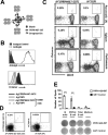
Figure 4. Antigen-specific B cell staining by the Ag7/PRPH467-507C octamer
(A) Schematic representation of the Ag7/PRPH467-507C octamer. PRPH467-507 is fused to the C-terminus of the α and ß chain of Ag7; the α chain is site-specifically labeled with biotin. SA, fluorescent streptavidin. (B) The B cell hybridoma I6 expressed surface IgG as detected through an IgG-specific Ab by FACS analysis (top). Staining of the same hybridoma by the Ag7/PRPH467-507C octamer, but not by the Ag7/GPI tetramer control molecule (bottom). Staining is lost if the Ag7/PRPH467-507C octamer is preincubated with the anti-PRPH mAb 228 E1 (right). (C) Detection of PRPHC B cells in pancreatic lymph nodes, spleen and peritoneum by Ag7/PRPH467-507C octamers. A FACS analysis of a 7 week-old NOD female is shown. B cells were gated on CD8, CD4, CD11c,F4/80, PI and CD19+. Left panels, staining with Ag7/PRPH467-507C octamers; right panels, staining with Ag7/GPI tetramers. (D) Detection of PRPHC B cells by FACS analysis by a 2-color staining approach in the peritoneum of a 7-wk-old NOD female. Total peritoneal cells were incubated with Ag7/PRPH467-507C octamers (left) or the control Ag7/GPI (right), both labeled with APC or PE. B cells are gated on CD8, CD4, CD11c, F4/80, PI and CD19+. (E) Ag-specificity of PRPHC B cells detected by ELISPOT analysis. 4000 PRPHC B cells, labeled using the 2-color approach as explained in (C) or 4000 CD19+ total B cells were sorted by FACS into ELISPOT plates, coated with PRPH492-507 or GST as background control. Cells were cultivated for 72 hrs in presence of anti-CD40 Abs, and secreted Ag-specific Abs were detected using an anti-mouse kappa Ab. As positive control, only 1000 I6 hybridomas cells were seeded in parallel in order to compensate for the stronger proliferative capacity of these cells. Data (mean ± SD) are representative of 2 independent experiments. The P value was determined by 2-way ANOVA test, ***P < 0.001.
Figure 5
Figure 5. PRPHC B cell distribution in NOD and C57BL/6 mice
(A) FACS analysis of PRPHC B cells in the peritoneum, spleen, and pancreatic lymph nodes (Pan LN) in NOD males and females (n=5-15/age group). PRPHC B cells are shown in the indicated organs and ages as percentage of total CD19+ B cells. Ag7/PRPH467-507C octamer+ cells were gated on CD3, CD11c, F4/80, PI and B220+. Values obtained with the Ag7/GPI control were subtracted during analysis. Error bar indicates mean values ± SD. The P value was determined by Mann Whitney t test. (B) Analysis of PRPHC B cells in the peritoneum of diabetic and pre-diabetic 15 week-old NOD/LtJ females (n=5). PRPHC B cells were analyzed as in (A). Left panel, PRPHC B cells are indicated as a percentage of total intraperitoneal B220+ B cells; right panel, total numbers of intraperitoneal PRPHC B cells of the same analysis. Error bar indicates mean values ± SD. The P value was determined by Mann Whitney t test. (C) Representative FACS profile of islet-infiltrating PRPHC B cells from 15 week-old NOD/LtJ females (a pooled analysis of 5 animals is shown). PRPHC B cells were analyzed as in (A). (D) Comparative analysis of PRPHC B cells in the peritoneum and spleen in 7 week-old NOD and C57BL/6 females (n=5). PRPHC B cells were analyzed as in (A). Error bar indicates mean values ± SD. The P value was determined by Mann Whitney t test.
Figure 6
Figure 6. PRPHC B cells segregate into different subpopulations and are activated in a tissue-specific fashion
(A) FACS analysis of PRPHC B cell subpopulations in the peritoneum, spleen and pancreatic lymph nodes of 7 week-old NOD females. PRPHC B cells were gated using the 2-color approach explained in Fig. 4 C and further analyzed using anti-CD5 and Mac-1 (top panels) or anti-IgM and anti-IgD Abs (bottom panels). Plots are representative of 3 independent experiments. (B) FACS analysis of islet-infiltrating PRPHC B cell phenotypes from 10 week-old NOD/LtJ females (a pooled analysis of 5 animals is shown). Analysis as in (A); Left panels, CD19+ total B cells, right panels, PRPHC B cells. Top, IgM vs. IgD, bottom CD5 vs. Mac-1 analysis. Plots are representative of 2 independent experiments. (C) Analysis of PRPHC B cell phenotypes in the spleen of 8 week-old NOD/LtJ females (n=5). PRPHC B cells were first quantified by the 2-color approach explained in Fig. 4 C and further analyzed using the CD23 and CD21 markers (FACS profiles not shown). Total B cells are shown for comparison. FO follicular B cells, MZ marginal zone B cells. Error bars indicate mean values ± SD. Values were not significantly different as determined by 1-way ANOVA test.

Similar articles

Cited by

References

    1. Favas C, Isenberg DA. B-cell-depletion therapy in SLE - what are the current prospects for its acceptance? Nat. Rev. Rheumatol. 2009;5:711–716. - PubMed
    1. Lehuen A, Diana J, Zaccone P, Cooke A. Immune cell crosstalk in type 1 diabetes. Nat. Rev. Immunol. 2010;10:501–513. - PubMed
    1. Wong FS, Wen L. B Cells in Autoimmune Diabetes. Rev. Diabet. Stud. 2005;2:121–135. - PMC - PubMed
    1. Serreze DV, Chapman HD, Varnum DS, Hanson MS, Reifsnyder PC, Richard SD, Fleming SA, Leiter EH, Shultz LD. B lymphocytes are essential for the initiation of T cell-mediated autoimmune diabetes: analysis of a new “speed congenic” stock of NOD.Ig mu null mice. J. Exp. Med. 1996;184:2049–2053. - PMC - PubMed
    1. Akashi T, Nagafuchi S, Anzai K, Kondo S, Kitamura D, Wakana S, Ono J, Kikuchi M, Niho Y, Watanabe T. Direct evidence for the contribution of B cells to the progression of insulitis and the development of diabetes in non-obese diabetic mice. Int. Immunol. 1997;9:1159–1164. - PubMed

Publication types

MeSH terms