. 2014 Apr 1;192(7):3166-79.
doi: 10.4049/jimmunol.1302617. Epub 2014 Mar 7.

B cells in T follicular helper cell development and function: separable roles in delivery of ICOS ligand and antigen

Affiliations

B cells in T follicular helper cell development and function: separable roles in delivery of ICOS ligand and antigen

Jason S Weinstein et al. J Immunol. .

Abstract

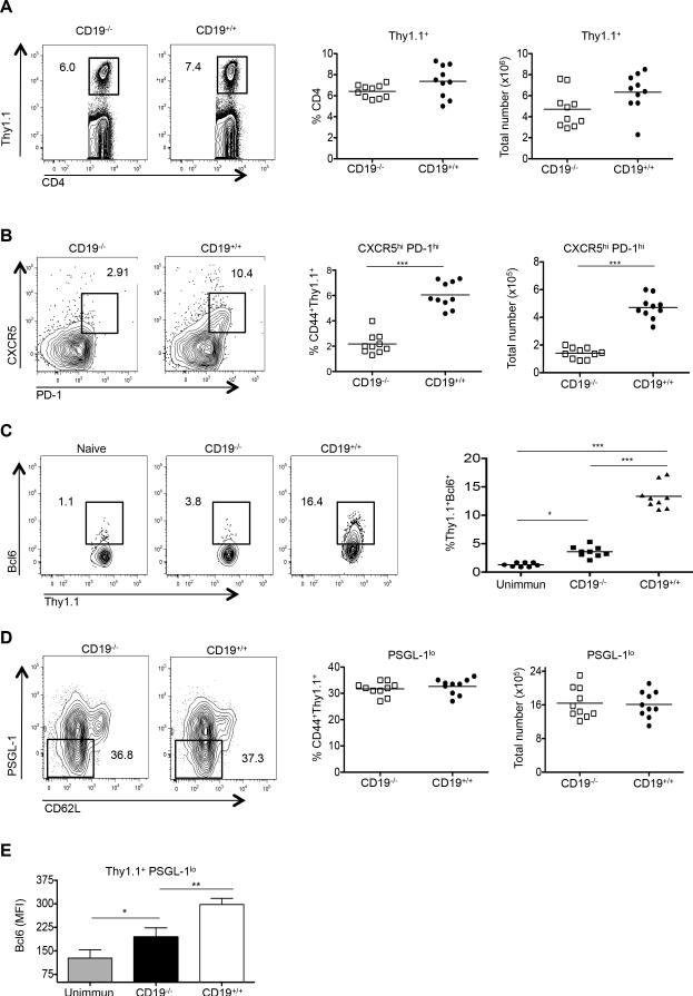
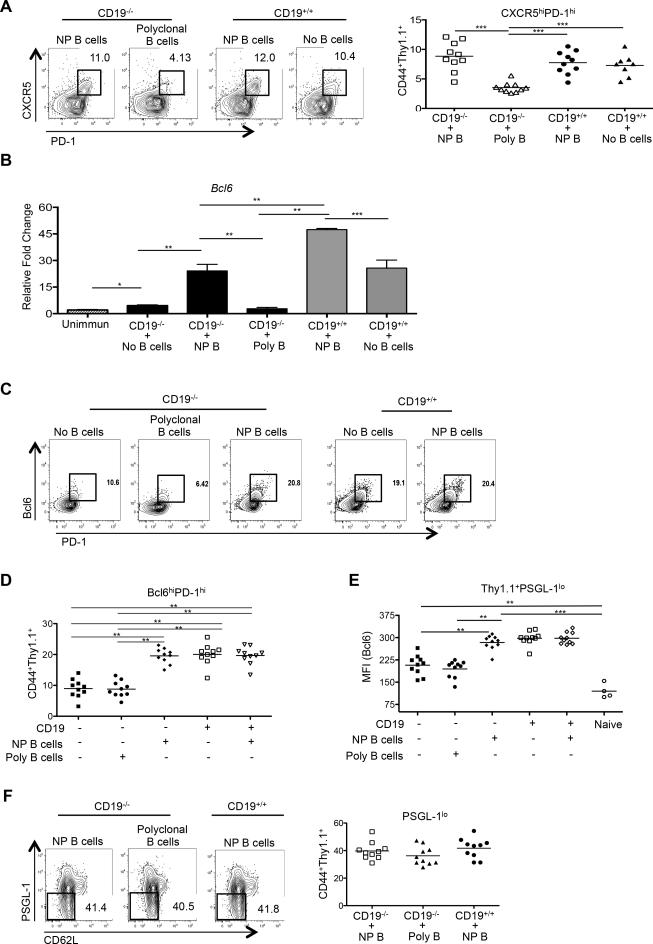
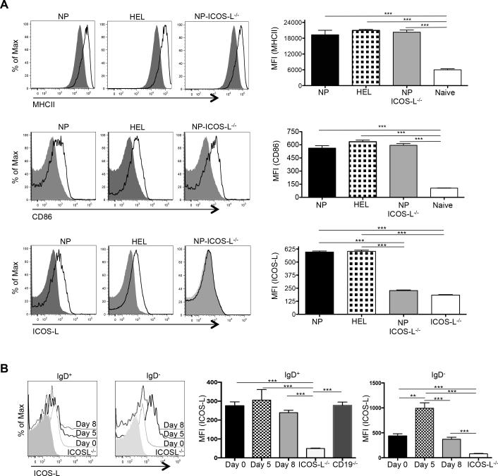
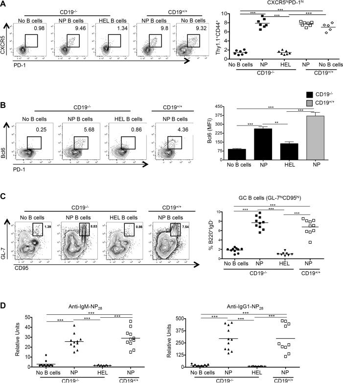
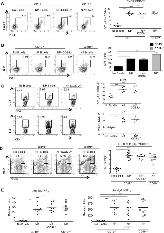
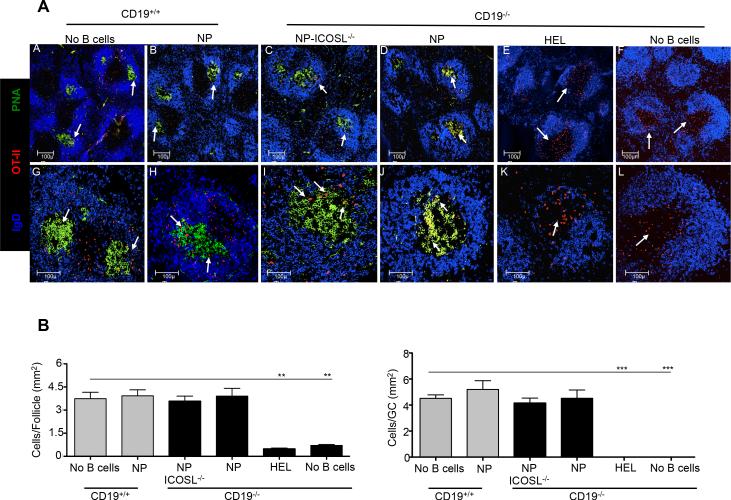
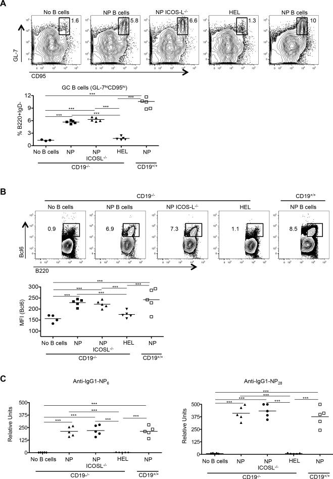
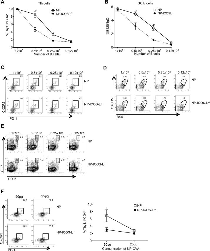
B cells are required for follicular Th (Tfh) cell development, as is the ICOS ligand (ICOS-L); however, the separable contributions of Ag and ICOS-L delivery by cognate B cells to Tfh cell development and function are unknown. We find that Tfh cell and germinal center differentiation are dependent on cognate B cell display of ICOS-L, but only when Ag presentation by the latter is limiting, with the requirement for B cell expression of ICOS-L overcome by robust Ag delivery. These findings demonstrate that Ag-specific B cells provide different, yet compensatory, signals for Tfh cell differentiation, while reconciling conflicting data indicating a requirement for ICOS-L expression on cognate B cells for Tfh cell development with those demonstrating that the latter requirement could be bypassed in lieu of that tendered by noncognate B cells. Our findings clarify the separable roles of delivery of Ag and ICOS-L by cognate B cells for Tfh cell maturation and function, and have implications for using therapeutic ICOS blockade in settings of abundantly available Ag, such as in systemic autoimmunity.


Figures

Figure 1
Figure 1
CD19-intact B cells are required for Tfh-cell development. CD19−/− (n = 10) or CD19+/+ (n = 10) mice received CD4+ Thy1.1+ OT-II TCR transgenic T cells, with spleens of recipients harvested 7 days after immunization with NP-OVA. (A) Representative flow cytometry plots of splenic cells showing the percentages of transferred Thy1.1 cells among total CD4 T cells (left 2 panels). The graphs (right 2 panels) show percentages and absolute numbers of CD4+ Thy1.1+ T cells following transfer to CD19−/− or CD19+/+ recipients. (B) Representative flow cytometry plots of splenic cells from CD19−/− or CD19+/+ recipients, as gated on CD4+ Thy1.1+CD44+ CXCR5hi PD-1hi T cells (left 2 panels). Graphs on the right show the percentages and total numbers of such cells. (C) Bcl6 expression in the transferred CD4+Thy1.1+ OT-II cells recovered from CD19−/−, CD19+/+, or unimmunized recipients (left 3 panels). Graph on the right shows the mean fluorescent intensity (MFI) of Bcl6 expression among the transferred CD4+ Thy1.1+ OT-II cells. (D) Representative PSGL-1 expression in the transferred CD4+ Thy1.1+ OT-II cells recovered from CD19−/− or CD19+/+ recipients (left 2 panels), with the percentages and numbers of these cells also shown (right 2 graphs). (E) Mean fluorescent intensity (MFI) of PSGL-1 expression among the transferred CD4+ Thy1.1+ OT-II cells, including into unimmunized recipients. All experiments were performed 3 times with n ≥ 5 mice per group. *** p < 0.001; ** p < 0.003; * p < 0.02 by Student's t-test comparing cells transferred into CD19−/− or CD19+/+ mice. Error bars represent standard deviation. Unimmun = unimmunized recipients.
Figure 2
Figure 2
Ag-specific B cells are required for antigen-specific Tfh-cell development. CD19−/− or CD19+/+ mice received CD4+ Thy1.1+ OT-II cells and NP-specific (n =10), polyclonal B cells (n = 10), or no B cells (n = 10), with spleens of recipients harvested 7 days after immunization with NP-OVA. (A) Representative flow cytometry plots of splenic cells from CD19−/− or CD19+/+ recipients, as gated on CD4+ Thy1.1+ CD44+ CXCR5hi PD-1hi T cells (left 3 panels), with the graph on the right showing the percentages of such cells among the transferred populations. (B) cDNA was synthesized from sorted CD4+ Thy1.1+ OT-II T cells from CD19−/− or CD19+/+ mice that received co-transfers of NP-specific or polyclonal B and CD4+ Thy1.1+ OT-II T cells. qPCR for Bcl6 was compared to that of Hprt. cDNA from sorted naïve CD4+ Thy1.1+ OT-II T cells (CD4+CD44lo) served as a control. (C and D) Representative Bcl6 and PD-1 expression on the transferred CD4+ T cells, including those transferred without B cells, in conjunction with numbers of such cells. (E) Bcl6 MFI from transferred CD4+ Thy1.1+ OT-II PSGL-1lo T cell populations. (F) Representative flow cytometry plots of splenic cells from CD19−/− or CD19+/+ recipients, as gated on CD4+Thy1.1+CD44+ PSGL-1lo T cells (left 3 panels), with the graph on the right showing the percentages of such cells. Experiments were performed 3 times with n ≥ 3 per group. ***p < 0.001; **p < 0.003; * p < 0.02 by Student's t-test comparing cells transferred into CD19−/− or CD19+/+ mice. Error bars represent standard deviation. Poly = polyclonal B cells.
Figure 3
Figure 3
Expression of surface proteins on adoptively transferred B cells. (A) Expression of class II MHC, CD86, and ICOS-L on NP-specific ICOS-L+/+ (n = 7), NP-ICOS-L−/− (n = 7) and HEL-specific MD4 B cells (n = 7) (solid black line) compared to expression of these proteins on naïve B cells (grey shaded area; left), with the MFI expression of these markers from each group of mice graphically displayed (right). (B) Expression of ICOS-L on transferred B cells before (day 0), and at days 5 and 8 after, immunization with NP-OVA (shaded histogram), with its MFI expression graphically displayed (right). ***p < 0.001; ** p < 0.003.
Figure 4
Figure 4
Ag-specific B cells are required for development of functional Tfh cells. CD19−/− or CD19+/+ mice received CD4 Thy1.1+ OT-II cells alone (n ≥ 5), or with NP-specific (n = 7) or HEL-specific MD4 B cells (n = 7), with spleens of recipients harvested 7 days after immunization with NP-OVA and sera collected. (A) Representative flow cytometry plots of CD4+ Thy1.1+ CD44+ CXCR5hi PD-1hi T cells from the recipients, with the graph on the right showing the percentages of such cells among the transferred population. (B) Representative Bcl6 and PD-1 expression in the transferred CD4+ T cells, including cells transferred without B cells (left 4 panels), in conjunction with Bcl6 expression (right panel). (C) Representative flow cytometry plots of B220+ IgD GL-7hi CD95hi GC B cells taken from the recipients, with the percentages of such cells among B220+ IgD cells shown on the graph on the right. (D) IgM (left) and IgG (right) anti-NP28 Ab levels in the recipients. Experiments were performed 3 times with n ≥ 3 per group. *** p < 0.001; ** p < 0.003 by Student's t-test comparing cells transferred into CD19−/− or CD19+/+ mice. Error bars represent standard deviation.
Figure 5
Figure 5
ICOS-L on Ag-specific B cells is dispensable for antigen-specific Tfh formation and function. CD19−/− or CD19+/+ mice received CD4 Thy1.1+ OT-II cells alone (n = 7-10), or with ICOS-L+/+ NP-specific (n = 7-10) or ICOS-L−/− NP-specific B cells (n = 7-10), with spleens of recipients harvested 7 days after immunization with NP-OVA and sera collected. (A) Representative flow cytometry plots of CD4+ Thy1.1+ CD44+ CXCR5hi PD-1hi T cells from the recipients, with the graph on the right showing the percentages of such cells among the transferred population. (B) Representative Bcl6 and PD-1 expression in the transferred CD4+ T cells, including cells transferred without B cells (left 4 panels), in conjunction with Bcl6 expression in the Thy1.1+ PSGL-1lo population in the various recipients (right panel). (C) Representative flow cytometry plots of IL-21 and IL-4 expression (top 3 and bottom 3 panels, respectively) in the CD4+ Thy1.1+ CD44+ CXCR5hi PD-1hi T cells from the various recipients, with aggregate totals of cytokine positive cells from Thy1.1+ PSGL-1lo population (right panels, n = 5 in each group). (D) Representative flow cytometry plots of B220+ IgD GL-7hi CD95hi GC B cells taken from the recipients, with the percentages of such cells among B220+ IgD cells shown on the graph on the right. (E) IgM (left) and IgG (right) anti-NP28 Ab levels in the recipients. Experiments were performed 3 times with n ≥ 3 per group. ***p < 0.001; **p < 0.003 by Student's t-test comparing cells transferred into CD19−/− or CD19+/+ mice. Error bars represent standard deviation.
Figure 6
Figure 6
Ag-specific B cells are required for follicular migration of antigen-specific T cells. CD19−/− or CD19+/+ mice received RFP+ CD4+ Thy1.1+ OT-II cells alone (n = 5), or with NP-specific (n = 5), NP-specific ICOS-L−/− (n = 5), or HEL-specific MD4 B cells (n = 5), with spleens of recipients harvested 7 days after immunization with NP-OVA followed by staining for confocal microscopy. (A) Representative follicles (10×) stained with anti-IgD (blue) or PNA (green), along with transferred RFP+ OT-II T cells (red) (panels A-F) or representative follicles, with the same stains, shown at higher magnification (25× objective) (panels G-L). Bars, 100 μm. (B) T cells localized within follicles (left) and GCs (right) were manually counted (blinded) using Image J software, with numbers of T cells per mm2 of follicle and GCs determined, using all the follicles and GCs examined. Grey and black bars = CD19+/+ and CD19−/− recipients, respectively. ** p < 0.03 and *** p = 0.01.
Figure 7
Figure 7
Cognate Tfh-B cell interactions are required for GC B cell development. CD19−/− or CD19+/+ mice received CD4+ Thy1.1+ OT-II cells alone (n = 5), or with NP-specific (n = 5), NP-specific ICOS-L−/− (n = 5), or HEL-specific MD4 B cells (n = 5), with spleens and sera of recipients harvested 14 days after immunization with NP-OVA. (A) Representative flow cytometry plots of B220+ IgD GL-7hi CD95hi GC B cells from CD19−/− or CD19+/+ recipients (top 5 panels), with their percentages of B220+ IgD cells (below). (B) Representative flow cytometry plots demonstrating the percentage of Bcl6+ B cells among the total B220+ IgD population in spleens of recipients (top 5 panels), with aggregate percentages of same (below). (C) High and low affinity anti-NP IgG1 Abs (anti-NP6 and anti-NP28; left and right panels, respectively) were determined from the different recipients. Experiments were performed 3 times with n ≥ 3 per group. ***p < 0.001; **p < 0.003; *p < 0.02 by Student's t-test comparing cells transferred into CD19−/− or CD19+/+ mice. Error bars represent standard deviation.
Figure 8
Figure 8
ICOS-L on B cells is required for Tfh-cell development and GC responses when Ag-presenting B cells or Ag are limited. (A) CD19−/− mice received 1 × 10⁶, 0.5 × 10⁶, 0.25 × 10⁶ or 0.12 × 10⁶ NP-specific or NP-specific ICOS-L−/− B cells co-transferred with 0.5 × 10⁶ CD4+ Thy1.1+ OT-II T cells, with spleens of recipients harvested 7 days after immunization with NP-OVA. Percentages of CD4+ Thy1.1+ CD44+ CXCR5hi PD-1hi T and B220+ IgD GL-7hi CD95hi B cells from animals receiving various numbers of NP-specific B cells are shown. (C-E) Representative flow cytometry plots of splenic CD4+ Thy1.1+ OT-II CD44+CXCR5hiPD-1hi or CD4+Thy1.1+CD44+CXCR5hiBcl6hi T cells, or B220+IgDloGL-7hiCD95hi B cells. Experiments were performed 3 times with n ≥ 5 per group. (F) CD4+ Thy1.1+ OT-II cells were transferred into CD19-deficient mice, followed by immunization with 100 μg NP-OVA. Two days after priming, 0.05 × 10⁶ OT-II T cells were re-transferred, along with 1 × 10⁶ ICOS-L-intact or –deficient NP-specific B cells, into CD19−/− animals primed 2 days earlier with 25 or 50 ug of NP-OVA, with spleens of recipients harvested 7 days after immunization. Representative flow cytometry plots of splenic CD4+ Thy1.1+ OT-II CD44+CXCR5hiPD-1hi Tfh cells with the percentages of such cells among CD4+ Thy1.1+ cells shown on the graph on the right. n = 3 mice per group. **p < 0.003; *p < 0.02 by Sidak's multiple comparison test comparing the means of Tfh (A and F) and GC B (B) cell percentages (of transferred cells) in recipient mice. Error bars represent standard deviation.

References

    1. King C, Tangye SG, Mackay CR. T follicular helper (TFH) cells in normal and dysregulated immune responses. Annu Rev Immunol. 2008;26:741–766. - PubMed
    1. Xu J, Foy TM, Laman JD, Elliott EA, Dunn JJ, Waldschmidt TJ, Elsemore J, Noelle RJ, Flavell RA. Mice deficient for the CD40 ligand. Immunity. 1994;1:423–431. - PubMed
    1. Zaretsky AG, Taylor JJ, King IL, Marshall FA, Mohrs M, Pearce EJ. T follicular helper cells differentiate from Th2 cells in response to helminth antigens. J Exp Med. 2009;206:991–999. - PMC - PubMed
    1. Zotos D, Coquet JM, Zhang Y, Light A, D'Costa K, Kallies A, Corcoran LM, Godfrey DI, Toellner KM, Smyth MJ, Nutt SL, Tarlinton DM. IL-21 regulates germinal center B cell differentiation and proliferation through a B cell-intrinsic mechanism. J Exp Med. 2010;207:365–378. - PMC - PubMed
    1. Linterman MA, Beaton L, Yu D, Ramiscal RR, Srivastava M, Hogan JJ, Verma NK, Smyth MJ, Rigby RJ, Vinuesa CG. IL-21 acts directly on B cells to regulate Bcl-6 expression and germinal center responses. J Exp Med. 2010;207:353–363. - PMC - PubMed

Publication types

MeSH terms