. 2014 May;196(10):1877-88.
doi: 10.1128/JB.01502-14. Epub 2014 Mar 7.

A novel ESX-1 locus reveals that surface-associated ESX-1 substrates mediate virulence in Mycobacterium marinum

Affiliations

A novel ESX-1 locus reveals that surface-associated ESX-1 substrates mediate virulence in Mycobacterium marinum

George M Kennedy et al. J Bacteriol. 2014 May.

Abstract

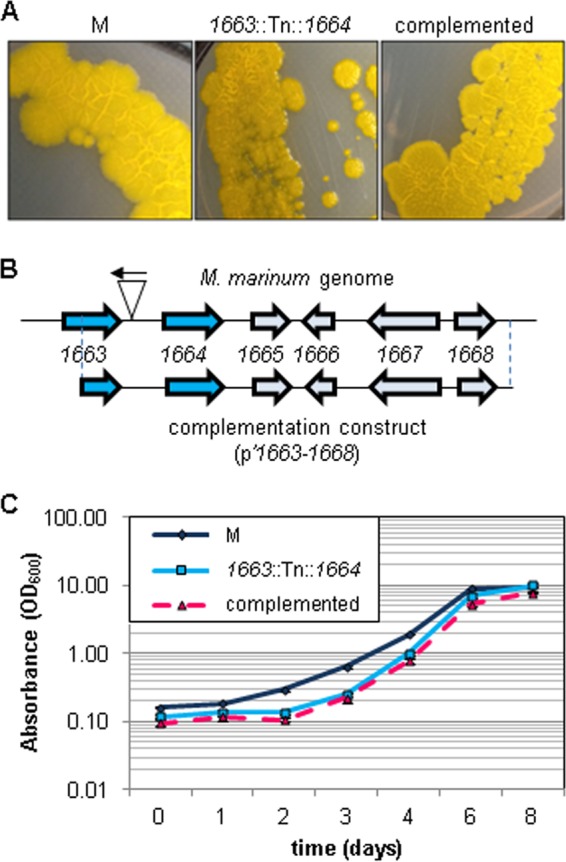
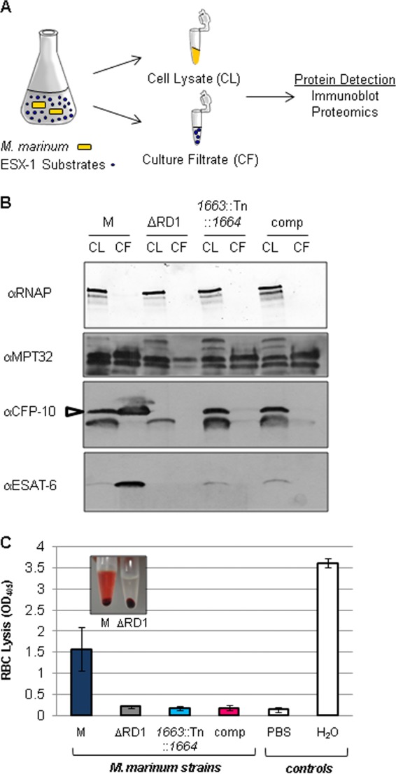
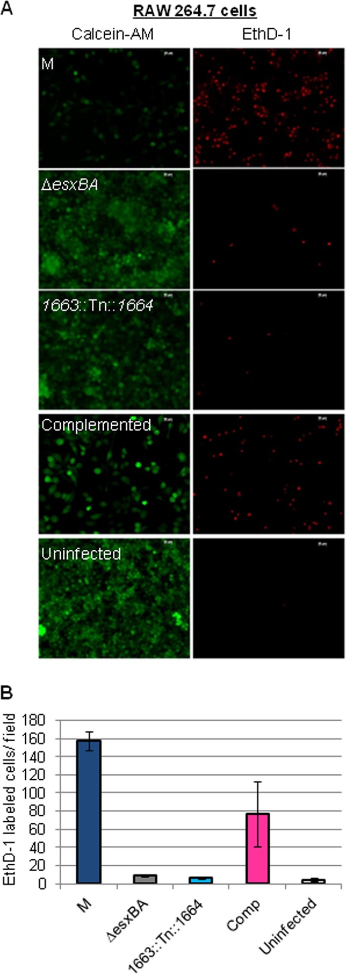
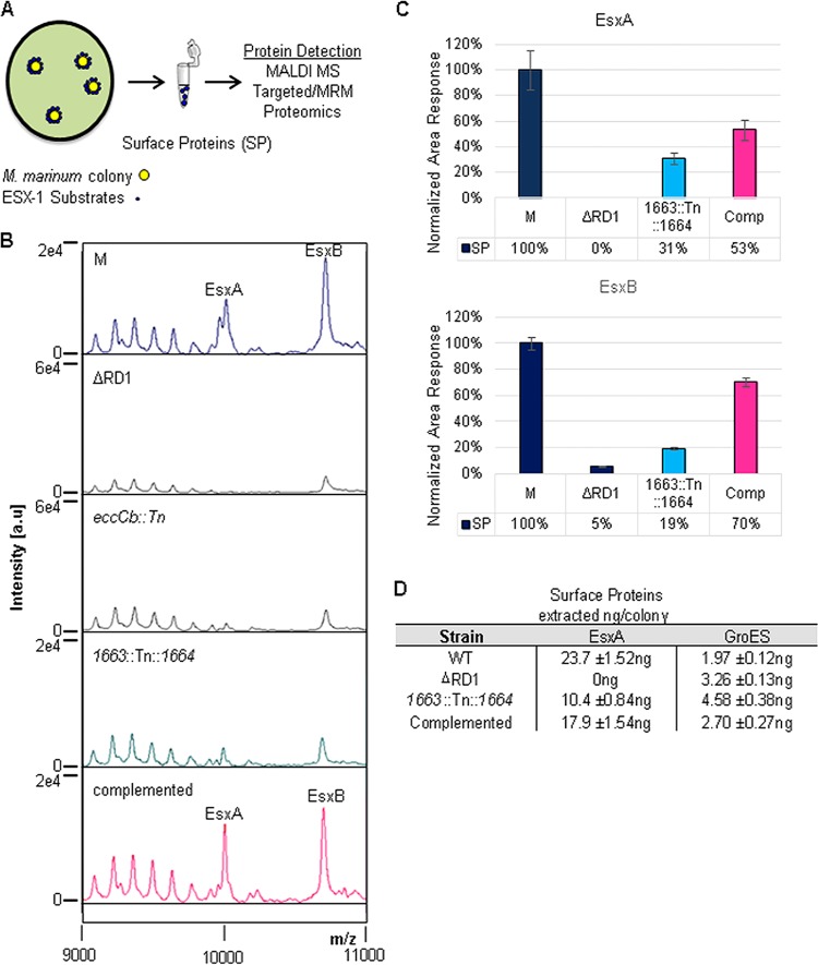
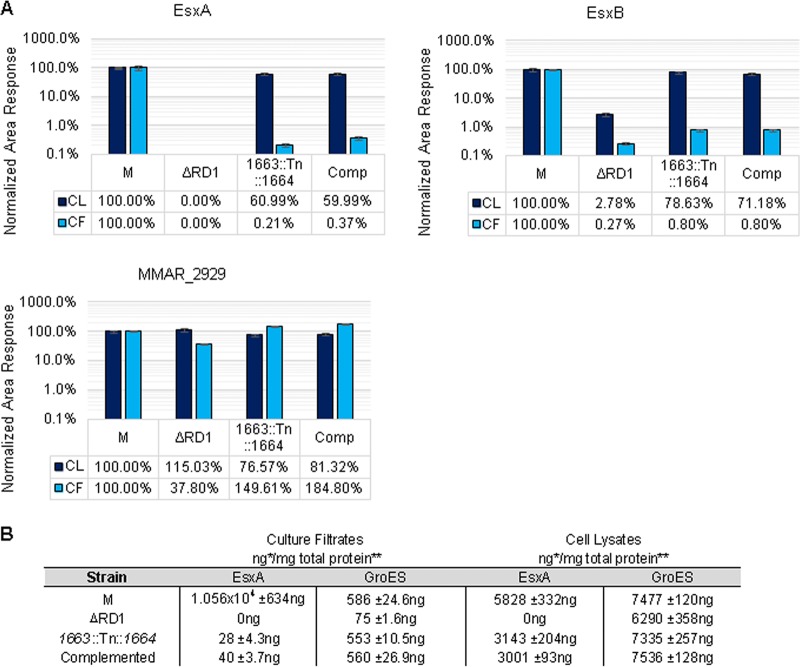
EsxA (ESAT-6) and EsxB (CFP-10) are virulence factors exported by the ESX-1 system in mycobacterial pathogens. In Mycobacterium marinum, an established model for ESX-1 secretion in Mycobacterium tuberculosis, genes required for ESX-1 export reside at the extended region of difference 1 (RD1) locus. In this study, a novel locus required for ESX-1 export in M. marinum was identified outside the RD1 locus. An M. marinum strain bearing a transposon-insertion between the MMAR_1663 and MMAR_1664 genes exhibited smooth-colony morphology, was deficient for ESX-1 export, was nonhemolytic, and was attenuated for virulence. Genetic complementation revealed a restoration of colony morphology and a partial restoration of virulence in cell culture models. Yet hemolysis and the export of ESX-1 substrates into the bacteriological medium in vitro as measured by both immunoblotting and quantitative proteomics were not restored. We show that genetic complementation of the transposon insertion strain partially restored the translocation of EsxA and EsxB to the mycobacterial cell surface. Our findings indicate that the export of EsxA and EsxB to the cell surface, rather than secretion into the bacteriological medium, correlates with virulence in M. marinum. Together, these findings not only expand the known genetic loci required for ESX-1 secretion in M. marinum but also provide an explanation for the observed disparity between in vitro ESX-1 export and virulence.


Figures

FIG 1
FIG 1
Identification and characterization of a smooth M. marinum strain. (A) Images of the indicated M. marinum strains grown on 7H11 agar plates showing the colony morphology phenotypes of the wild-type M strain, the Tn insertion strain, and the complemented strain. (B) The site of Tn insertion in the M. marinum strain 14 genome. The Tn inserted 120 bp upstream of the MMAR_1664 gene. The construct used for complementation is indicated below the locus and included the C-terminal half of the MMAR_1663 gene through the MMAR_1668 gene. (C) Growth as measured by absorbance at the OD600 of M. marinum cultures in 7H9 defined broth. Error bars represent standard deviations.
FIG 2
FIG 2
MMAR_1663-MMAR_1668 is required for ESX-1 export. (A) Schematic of the in vitro ESX-1 secretion assay. (B) M. marinum ESX-1 substrate production and secretion into the bacteriological medium as detected by Western blot analysis. The RNAP beta subunit served as a control for lysis and as a loading control for the cell lysate (CL); MPT-32 served as loading control for the culture filtrate (CF). The CFP-10 antibody detected the EsxB protein from M. marinum (indicated by the open arrow). A cross-reacting band is present below the EsxB band as determined by the lack of the top band in the ΔRD1 control strain. The ESAT-6 antibody detected the EsxA protein from M. marinum. The ΔRD1 strain served as a control for antibody specificity. α, anti; comp, complemented strain. (C) Hemolysis assay to measure ESX-1 function. PBS served as a negative control, and water served as a positive control for RBC lysis. Error bars represent the standard deviations. Representative experiments of three biological replicates are shown. The inset is a photograph of the hemolysis assay for the M and ΔRD1 strains.
FIG 3
FIG 3
The MMAR_1663-MMAR_1668 region of the M. marinum genome is required for virulence. M. marinum infection of the RAW 264.7 cell line was performed at an MOI of 10. Images were acquired at 24 h postinfection with a 20× objective on a Zeiss Axio Observer microscope. Scale bar, 20 mm. EthD-1 staining showed membrane permeabilization of the macrophages, resulting in red fluorescence. Calcein-AM labeled live macrophages following infection. (B) Quantitation of the infection data shown in panel A. Red EthD-1-stained cells per field were counted; four independent fields from each infection were counted, and the counts were averaged. Error bars indicate the standard deviations between fields.
FIG 4
FIG 4
Complementation partially restores the translocation of EsxA and EsxB to the mycobacterial cell surface. (A) Schematic of the whole-colony MALDI MS ESX-1 secretion assay. (B) MALDI peaks for EsxA and EsxB generated from surface proteins from M. marinum strains. The ΔRD1 strain lacks the genes encoding EsxA and EsxB and served as a control for specificity. The eccCb::Tn strain produces but does not export EsxA or EsxB and served as a control for lysis. (C) Differential quantitative mass spectrometry-based analysis of surface protein (SP) complementation from the wild-type, ΔRD1, mutant, and complemented strains. Relative quantification was determined by analysis of tryptic digests of washed colony extracts (surface proteins) compared using LC-MS/MS (MRM-based) measurement. Lysis and normalization were performed using levels of GroES and GroEL. The ΔRD1 strain served as a negative ESX-1 and lysis control. Standard error is shown from triplicate analysis. (D) Absolute levels of EsxA and GroES from colony surface proteins from the wild-type (WT), ΔRD1, 1663::Tn::1664, and complemented strains. Quantification was determined by LC-MS/MS (MRM-based) analysis of tryptic digests with the addition of 13C15N stable heavy-isotope versions of peptides from each protein. EsxA values shown are normalized to the GroES values shown. Standard error is shown from triplicate analyses. Large colonies yielded about 10 to 20 μg of total soluble protein.
FIG 5
FIG 5
Differential and absolute quantification of ESX-1 substrates in M. marinum. Relative levels of EsxA, EsxB, and MMAR_2929 in whole-cell lysates and culture supernatants from the ΔRD1, 1663::Tn::1664, and complemented M. marinum strains were compared to levels of the wild-type M strain. Error bars represent the average percent coefficient of variation of the cell lysates (CL) and culture filtrates (CF). (B) Absolute levels of EsxA and GroES in whole-cell lysates and culture supernatants generated from the wild-type, ΔRD1, 1663::Tn::1664, and complemented strains. Quantification was determined by analyzing tryptic digests by LC-MS/MS triple-quadrupole MRM-based proteomics with 13C15N stable heavy-isotope versions of peptides from each protein. Our linear dynamic range was 4 orders of magnitude, and the LLOQs were approximately 2.5 ng/mg for EsxA and 250 pg/mg for GroES. Error shown is the percent coefficient of variation of the measured value. *, MRM value; **, bicinchoninic acid protein assay of bulk material.

References

    1. Burts ML, Williams WA, DeBord K, Missiakas DM. 2005. EsxA and EsxB are secreted by an ESAT-6-like system that is required for the pathogenesis of Staphylococcus aureus infections. Proc. Natl. Acad. Sci. U. S. A. 102:1169–1174. 10.1073/pnas.0405620102 - DOI - PMC - PubMed
    1. Garufi G, Butler E, Missiakas D. 2008. ESAT-6-like protein secretion in Bacillus anthracis. J. Bacteriol. 190:7004–7011. 10.1128/JB.00458-08 - DOI - PMC - PubMed
    1. Guinn KM, Hickey MJ, Mathur SK, Zakel KL, Grotzke JE, Lewinsohn DM, Smith S, Sherman DR. 2004. Individual RD1-region genes are required for export of ESAT-6/CFP-10 and for virulence of Mycobacterium tuberculosis. Mol. Microbiol. 51:359–370. 10.1046/j.1365-2958.2003.03844.x - DOI - PMC - PubMed
    1. Hsu T, Hingley-Wilson SM, Chen B, Chen M, Dai AZ, Morin PM, Marks CB, Padiyar J, Goulding C, Gingery M, Eisenberg D, Russell RG, Derrick SC, Collins FM, Morris SL, King CH, Jacobs WR., Jr 2003. The primary mechanism of attenuation of bacillus Calmette-Guerin is a loss of secreted lytic function required for invasion of lung interstitial tissue. Proc. Natl. Acad. Sci. U. S. A. 100:12420–12425. 10.1073/pnas.1635213100 - DOI - PMC - PubMed
    1. Stanley SA, Raghavan S, Hwang WW, Cox JS. 2003. Acute infection and macrophage subversion by Mycobacterium tuberculosis require a specialized secretion system. Proc. Natl. Acad. Sci. U. S. A. 100:13001–13006. 10.1073/pnas.2235593100 - DOI - PMC - PubMed

Publication types

MeSH terms