Hematopoietic microRNA-126 protects against renal ischemia/reperfusion injury by promoting vascular integrity
- PMID: 24610930
- PMCID: PMC4116053
- DOI: 10.1681/ASN.2013060640
Hematopoietic microRNA-126 protects against renal ischemia/reperfusion injury by promoting vascular integrity
Abstract
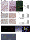
Ischemia/reperfusion injury (IRI) is a central phenomenon in kidney transplantation and AKI. Integrity of the renal peritubular capillary network is an important limiting factor in the recovery from IRI. MicroRNA-126 (miR-126) facilitates vascular regeneration by functioning as an angiomiR and by modulating mobilization of hematopoietic stem/progenitor cells. We hypothesized that overexpression of miR-126 in the hematopoietic compartment could protect the kidney against IRI via preservation of microvascular integrity. Here, we demonstrate that hematopoietic overexpression of miR-126 increases neovascularization of subcutaneously implanted Matrigel plugs in mice. After renal IRI, mice overexpressing miR-126 displayed a marked decrease in urea levels, weight loss, fibrotic markers, and injury markers (such as kidney injury molecule-1 and neutrophil gelatinase-associated lipocalin). This protective effect was associated with a higher density of the peritubular capillary network in the corticomedullary junction and increased numbers of bone marrow-derived endothelial cells. Hematopoietic overexpression of miR-126 increased the number of circulating Lin(-)/Sca-1(+)/cKit(+) hematopoietic stem and progenitor cells. Additionally, miR-126 overexpression attenuated expression of the chemokine receptor CXCR4 on Lin(-)/Sca-1(+)/cKit(+) cells in the bone marrow and increased renal expression of its ligand stromal cell-derived factor 1, thus favoring mobilization of Lin(-)/Sca-1(+)/cKit(+) cells toward the kidney. Taken together, these results suggest overexpression of miR-126 in the hematopoietic compartment is associated with stromal cell-derived factor 1/CXCR4-dependent vasculogenic progenitor cell mobilization and promotes vascular integrity and supports recovery of the kidney after IRI.
Copyright © 2014 by the American Society of Nephrology.
Figures


Comment in
-
Stem cells: The renoprotective role of miR-126.Nat Rev Nephrol. 2014 May;10(5):242. doi: 10.1038/nrneph.2014.52. Epub 2014 Mar 25. Nat Rev Nephrol. 2014. PMID: 24662430 No abstract available.
Similar articles
-
Silencing of miRNA-126 in kidney ischemia reperfusion is associated with elevated SDF-1 levels and mobilization of Sca-1+/Lin- progenitor cells.Microrna. 2014;3(3):144-9. doi: 10.2174/2211536604666150121000340. Microrna. 2014. PMID: 25541911
-
Postconditioning attenuates renal ischemia-reperfusion injury by mobilization of stem cells.J Nephrol. 2015 Jun;28(3):289-98. doi: 10.1007/s40620-015-0171-7. Epub 2015 Feb 6. J Nephrol. 2015. PMID: 25663348
-
SDF-1 provides morphological and functional protection against renal ischaemia/reperfusion injury.Nephrol Dial Transplant. 2010 Dec;25(12):3852-9. doi: 10.1093/ndt/gfq311. Epub 2010 Jun 2. Nephrol Dial Transplant. 2010. PMID: 20519232
-
Contribution of endothelial progenitors and proangiogenic hematopoietic cells to vascularization of tumor and ischemic tissue.Curr Opin Hematol. 2006 May;13(3):175-81. doi: 10.1097/01.moh.0000219664.26528.da. Curr Opin Hematol. 2006. PMID: 16567962 Free PMC article. Review.
-
miR-21 in ischemia/reperfusion injury: a double-edged sword?Physiol Genomics. 2014 Nov 1;46(21):789-97. doi: 10.1152/physiolgenomics.00020.2014. Epub 2014 Aug 26. Physiol Genomics. 2014. PMID: 25159851 Free PMC article. Review.
Cited by
-
Induction of microRNA-17-5p by p53 protects against renal ischemia-reperfusion injury by targeting death receptor 6.Kidney Int. 2017 Jan;91(1):106-118. doi: 10.1016/j.kint.2016.07.017. Epub 2016 Sep 9. Kidney Int. 2017. PMID: 27622990 Free PMC article.
-
Kidney Injury: Focus on Molecular Signaling Pathways.Curr Med Chem. 2024;31(28):4510-4533. doi: 10.2174/0109298673271547231108060805. Curr Med Chem. 2024. PMID: 38314680 Review.
-
The link between gestational diabetes and cardiovascular diseases: potential role of extracellular vesicles.Cardiovasc Diabetol. 2022 Sep 3;21(1):174. doi: 10.1186/s12933-022-01597-3. Cardiovasc Diabetol. 2022. PMID: 36057662 Free PMC article. Review.
-
The Circular RNA ciRs-126 Predicts Survival in Critically Ill Patients With Acute Kidney Injury.Kidney Int Rep. 2018 Jun 2;3(5):1144-1152. doi: 10.1016/j.ekir.2018.05.012. eCollection 2018 Sep. Kidney Int Rep. 2018. PMID: 30197981 Free PMC article.
-
MicroRNA-126 protects against vascular injury by promoting homing and maintaining stemness of late outgrowth endothelial progenitor cells.Stem Cell Res Ther. 2020 Jan 21;11(1):28. doi: 10.1186/s13287-020-1554-9. Stem Cell Res Ther. 2020. PMID: 31964421 Free PMC article.
References
-
- Perico N, Cattaneo D, Sayegh MH, Remuzzi G: Delayed graft function in kidney transplantation. Lancet 364: 1814–1827, 2004 - PubMed
-
- Pagtalunan ME, Olson JL, Tilney NL, Meyer TW: Late consequences of acute ischemic injury to a solitary kidney. J Am Soc Nephrol 10: 366–373, 1999 - PubMed
-
- Sharfuddin AA, Molitoris BA: Pathophysiology of ischemic acute kidney injury. Nat Rev Nephrol 7: 189–200, 2011 - PubMed
Publication types
MeSH terms
Substances
LinkOut - more resources
Full Text Sources
Other Literature Sources
Medical
Research Materials

