. 2014 May 1;306(9):L840-54.
doi: 10.1152/ajplung.00155.2013. Epub 2014 Mar 7.

Cigarette smoke-induced mitochondrial fragmentation and dysfunction in human airway smooth muscle

Affiliations

Cigarette smoke-induced mitochondrial fragmentation and dysfunction in human airway smooth muscle

Bharathi Aravamudan et al. Am J Physiol Lung Cell Mol Physiol. .

Abstract

The balance between mitochondrial fission and fusion is crucial for mitochondria to perform its normal cellular functions. We hypothesized that cigarette smoke (CS) disrupts this balance and enhances mitochondrial dysfunction in the airway. In nonasthmatic human airway smooth muscle (ASM) cells, CS extract (CSE) induced mitochondrial fragmentation and damages their networked morphology in a concentration-dependent fashion, via increased expression of mitochondrial fission protein dynamin-related protein 1 (Drp1) and decreased fusion protein mitofusin (Mfn) 2. CSE effects on Drp1 vs. Mfn2 and mitochondrial network morphology involved reactive oxygen species (ROS), activation of extracellular signal-regulated kinase (ERK), phosphatidylinositol 3-kinase (PI3K)/protein kinase B (Akt), protein kinase C (PKC) and proteasome pathways, as well as transcriptional regulation via factors such as NF-κB and nuclear erythroid 2-related factor 2. Inhibiting Drp1 prevented CSE effects on mitochondrial networks and ROS generation, whereas blocking Mfn2 had the opposite, detrimental effect. In ASM from asmatic patients, mitochondria exhibited substantial morphological defects at baseline and showed increased Drp1 but decreased Mfn2 expression, with exacerbating effects of CSE. Overall, these results highlight the importance of mitochondrial networks and their regulation in the context of cellular changes induced by insults such as inflammation (as in asthma) or CS. Altered mitochondrial fission/fusion proteins have a further potential to influence parameters such as ROS and cell proliferation and apoptosis relevant to airway diseases.

Keywords: asthma; dynamin-related protein 1; lung; mitochondria; mitofusin 2; signaling.


Figures

Fig. 1.
Fig. 1.
Changes in mitochondrial morphology of human airway smooth muscle (ASM) cells caused by cigarette smoke (CS). A: representative images of MitoTracker Green-labeled ASM mitochondria upon exposure to 0 (untreated), 0.5, 1, or 2% cigarette smoke extract (CSE) for 24 or 48 h. B: representative scatter plot showing the range of mitochondrial morphology parameters (form factor representing mitochondrial branching and aspect ratio presenting branch length) at baseline and upon 1% CSE treatment. Each blue or red circle represents individual cells from one patient of control vs. 1% CSE group, respectively. C and D: quantification of changes in mitochondrial morphology in response to different concentrations of CSE showed concentration-dependent decreases in form factor (C) and aspect ratio (D) compared with untreated cells at both 24 and 48 h of CSE exposure. *Significant effect of CSE compared with untreated control. %Significant CSE concentration dependence (P < 0.05).
Fig. 2.
Fig. 2.
Effect of CSE on mitochondrial fission and fusion proteins in human ASM. A: in human ASM cells, exposure to 1% CSE increased mRNA expression for the fission protein dynamin-related protein (Drp1) but decreased mRNA levels for the fusion mitofusins (Mfn1, Mfn2). B: these mRNA changes were matched by increased expression of Drp1 and decreased expression of Mfn2 protein in human ASM cell lysates. C and D: quantification of the immunoblots showed that exposure to different concentrations of CSE for either 24 (C) or 48 (D) h resulted in concentration-dependent changes in Drp1 vs. Mfn2. *Significant effect of CSE compared with untreated control. %Significant CSE concentration dependence (P < 0.05).
Fig. 3.
Fig. 3.
Role of mitochondrial fission and fusion proteins in mediating effects of CSE on human ASM. A: immunoblot showing inhibition efficiency of Mfn2 and Drp1 small-interfering RNAs (siRNAs) with absence of effect of nontargeting RNA. GAPDH was used as a loading control. B: densitometric quantification of immunoblot data showing Mfn2 and Drp1 siRNA efficacy. C and D: quantification of mitochondrial morphology, in terms of form factor (C) and aspect ratio (D), showed that both morphological parameters are increased when Drp1 expression is suppressed (consistent with a fission role for Drp1), whereas parameters are reduced when Mfn2 expression is inhibited (consistent with a fusion role for Mfn2). siRNAs against either protein substantially reversed changes in mitochondrial morphology induced by 1% CSE, albeit to different extents. *Significant effect of CSE compared with untreated control. #Significant siRNA effect (P < 0.05).
Fig. 4.
Fig. 4.
Multiple signaling pathways mediate CSE effects on mitochondrial fission and fusion proteins in human ASM. A and B: quantification of mitochondrial morphology, in terms of form factor (A) and aspect ratio (B), when ASM cells are exposed to 1% CSE, in the presence of inhibitors for mitogen-activated protein (MAP) kinase (PD-98059), protein kinase B (Akt) (Akt inhibitor XIII), phosphatidylinositol 3-kinase (PI3K) (wortmannin), protein kinase C (PKC) (bisindolylmaleimide; BIS), or proteasome (lactacystin). Perturbing these pathways blocked CSE-induced mitochondrial fragmentation, albeit to different extents, with substantial effect of lactacystin and PD-98059, particularly for form factor. C and D: these morphological changes were accompanied by appropriate lack of changes in expression of Drp1 vs. Mfn2 following 1% CSE exposure. *Significant effect of CSE compared with untreated control. $Significant inhibitor effect (P < 0.05).
Fig. 5.
Fig. 5.
Cytosol-to-nuclear signals mediate CSE effects on mitochondrial fission and fusion in human ASM. A and B: quantification of mitochondrial morphology, in terms of form factor (A) and aspect ratio (B), when ASM cells are exposed to 1% CSE, in the presence of inhibitors of NF-κB (SN50) or nuclear erythroid 2-related factor 2 (Nrf2, trigonelline). Perturbing these nuclear factors blunted CSE-induced mitochondrial fragmentation, albeit to different extents. C and D: these morphological changes were reflected by blunted effects of CSE on Drp1 vs. Mnf2. *Significant effect of CSE compared with untreated control. $Significant inhibitor effect (P < 0.05).
Fig. 6.
Fig. 6.
Mitochondrial fission and fusion apparatus drives activation of transcription factors NF-κB and Nrf2. A: representative immunoblots showing nuclear translocation of NF-κB subunits P50 and P65, and of Nrf2, following 1% CSE exposure in vehicle-transfected control ASM cells vs. those transfected with Drp1 vs. Mfn2 siRNA. Whereas inhibiting Drp1 did not have any effect on translocation in control cells, inhibiting Mfn2 enhanced such translocation (A, BD show summaries). Furthermore, Drp1 siRNA blunted CSE-induced translocation of nuclear factors, whereas Mfn2 siRNA enhanced it. Nontargeting RNA was without effect. C, cytoplasmic fraction; N, nuclear fraction. *Significant effect of CSE compared with untreated control. #Significant siRNA effect (P < 0.05).
Fig. 7.
Fig. 7.
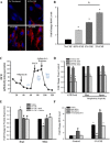
CSE, mitochondrial fission and fusion proteins, and reactive oxygen species (ROS) in human ASM. A: levels of ROS in ASM mitochondria following CSE treatment were determined using MitoSOX Red dye (DAPI for nuclear stains post hoc). B: quantification of MitoSOX Red fluorescence showed that exposure to CSE resulted in a concentration-dependent increase in ROS levels. C and D: the adequacy of endogenous antioxidants in these observations was verified using measurements of mitochondrial respiration parameters via a Seahorse Bioanalyzer. C shows representative tracings of oxygen consumption rate following different interventions to elicit parameters such as ATP production and maximum and spare respiratory capacities (summarized in D). OCR, oxygen consumption rate. Addition of artificial epithelial lining fluid (ELF) did not substantially influence OCR or other respiratory parameters in control cells or with the addition of 1% CSE. E: introduction of antioxidants restored mitochondrial fission-fusion balance by inhibiting CSE effects on human ASM. F: conversely, Drp1 siRNA prevented CSE-induced elevation in ROS levels, whereas Mfn2 siRNA enhanced it. *Significant effect of CSE compared with untreated control. %CSE concentration dependence. #Significant siRNA effect (P < 0.05).
Fig. 8.
Fig. 8.
Effect of CSE on mitochondrial morphology and ROS in human ASM from moderate asmatic patients. A and B: ASM cells from asmatic subjects showed baseline increased fragmentation reflected by form factor (A) and aspect ratio (B) such that subsequent CSE have less effect. C: on the other hand, mRNA and protein analysis suggested that baseline expression of fission proteins (Drp1, Fis1) were increased in ASM from asmatic patients, whereas Mfn1 and -2 were reduced. Importantly, exposure to 1% CSE exacerbated these changes. D: consistent with these morphological changes, Drp1 siRNA blunted CSE-induced elevation in ROS. ASM cells from three asthmatic vs. nonasthmatic individuals each were used for these assays. @Significant difference between asmatic subjects vs. nonasmatic subjects. *Significant effect of CSE compared with untreated control. #Significant siRNA effect (P < 0.05).
Fig. 9.
Fig. 9.
Schematic showing the role of mitochondrial proteins in mediating and modulating CS effects in human ASM. Under normal conditions, Mfn2 helps maintain mitochondrial networks with elongated branching (fusion), whereas the role of Drp1, which promotes fragmentation, is less. With CS exposure (or concomitant inflammation in the case of asthma), Mfn2 expression and function is reduced, whereas Drp1-mediated mitochondrial fragmentation increases. These processes are mediated and modulated by ROS and by a range of signaling pathways relevant to airway inflammation. In turn, Drp1 and Mfn2 can modulate these pathways. These interactions can overall lead to more fragmented mitochondrial networks with downstream consequences such as elevated ROS, cell proliferation vs. apoptosis, etc.

Comment in

References

    1. Aguilera-Aguirre L, Bacsi A, Saavedra-Molina A, Kurosky A, Sur S, Boldogh I. Mitochondrial dysfunction increases allergic airway inflammation. J Immunol 183: 5379–5387, 2009 - PMC - PubMed
    1. Alirol E, James D, Huber D, Marchetto A, Vergani L, Martinou JC, Scorrano L. The mitochondrial fission protein hFis1 requires the endoplasmic reticulum gateway to induce apoptosis. Mol Biol Cell 17: 4593–4605, 2006 - PMC - PubMed
    1. Archer SL, Gomberg-Maitland M, Maitland ML, Rich S, Garcia JG, Weir EK. Mitochondrial metabolism, redox signaling, and fusion: a mitochondria-ROS-HIF-1alpha-Kv1.5 O2-sensing pathway at the intersection of pulmonary hypertension and cancer. Am J Physiol Heart Circ Physiol 294: H570–H578, 2008 - PubMed
    1. Baglole CJ, Bushinsky SM, Garcia TM, Kode A, Rahman I, Sime PJ, Phipps RP. Differential induction of apoptosis by cigarette smoke extract in primary human lung fibroblast strains: implications for emphysema. Am J Physiol Lung Cell Mol Physiol 291: L19–L29, 2006 - PubMed
    1. Bargagli E, Olivieri C, Bennett D, Prasse A, Muller-Quernheim J, Rottoli P. Oxidative stress in the pathogenesis of diffuse lung diseases: a review. Respir Med 103: 1245–1256, 2009 - PubMed

Publication types

MeSH terms