. 2013 Dec 28;46(6):171-7.
doi: 10.1267/ahc.13035. Epub 2013 Dec 25.

Distribution of podoplanin-expressing cells in the mouse nervous systems

Affiliations

Distribution of podoplanin-expressing cells in the mouse nervous systems

Miwa Tomooka et al. Acta Histochem Cytochem. .

Abstract

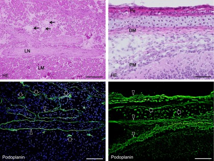
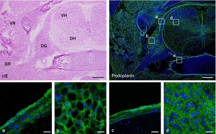
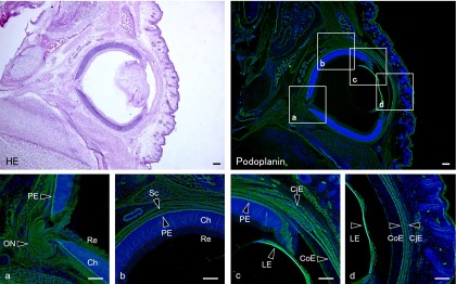
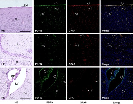
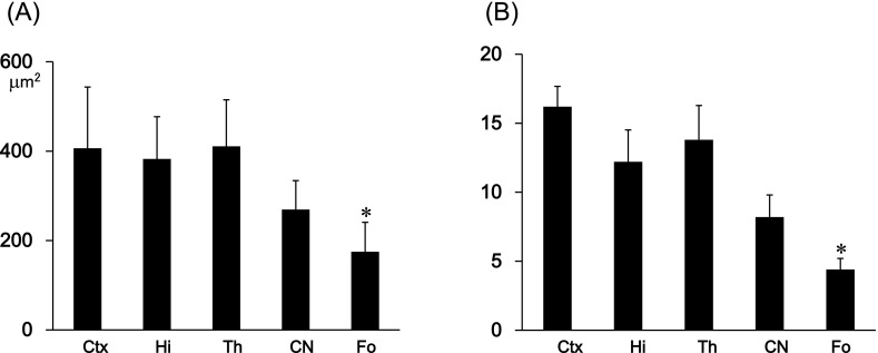
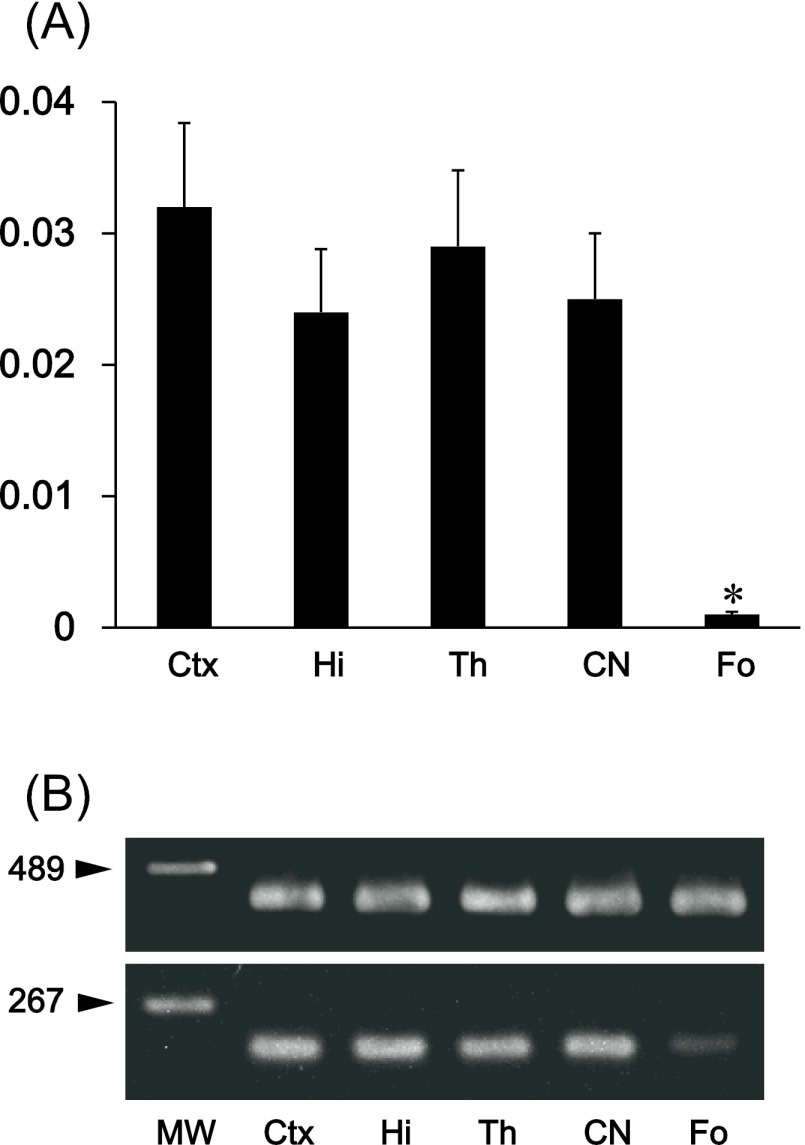
Podoplanin is a mucin-type glycoprotein which was first identified in podocytes. Recently, podoplanin has been successively reported as a marker for brain and peripheral nerve tumors, however, the distribution of podoplanin-expressing cells in normal nerves has not been fully investigated. This study aims to examine the podoplanin-expressing cell distribution in the mouse head and nervous systems. An immunohistochemical study showed that the podoplanin-positive areas in the mouse peripheral nerve and spinal cord are perineurial fibroblasts, satellite cells in the dorsal root ganglion, glia cells in the ventral and dorsal horns, and schwann cells in the ventral and dorsal roots; in the cranial meninges the dura mater, arachnoid, and pia mater; in the eye the optic nerve, retinal pigment epithelium, chorioidea, sclera, iris, lens epithelium, corneal epithelium, and conjunctival epithelium. In the mouse brain choroid plexus and ependyma were podoplanin-positive, and there were podoplanin-expressing brain parenchymal cells in the nuclei and cortex. The podoplanin-expressing cells were astrocyte marker GFAP-positive and there were no differences in the double positive cell distribution of several portions in the brain parenchyma except for the fornix. The results suggest that podoplanin may play a common role in nervous system support cells and eye constituents.

Keywords: astrocyte; glia cell; nervous system; podoplanin.


Figures

Fig. 1
Fig. 1
Podoplanin expression in mouse lymphatic vessels, bone, and connective tissue. The immunostained sections were re-stained by H-E staining. In the mouse tongue, the lingual muscle and blood vessels (arrows in HE) are podoplanin-negative but lymphatic vessels (arrows in immunostaining) and the perineurium around the lingual nerve (arrowheads) are podoplanin-positive. In the cranium, podoplanin-positive areas (arrowheads) are observed in periosteum (Pe), dura mater (DM), and pia mater (PM). Osteocytes in the cranial bone are also podoplanin-positive (arrows). Bar=100 µm.
Fig. 2
Fig. 2
Immunostaining for podoplanin in a horizontal section of mouse spinal cord. The immunostained sections were re-stained by H-E staining. The regions which are highlighted by boxes in the top right podoplanin immunostaining panel correspond to the lower panels with laser-scanning microscopic images (a–d). In the spinal cord, the podoplanin-positive areas are observed in (a) meninges, (b) satellite cells of the dorsal root ganglion (DG), (c) periosteum of the vertebra, and (d) glia cells of the ventral horn (VH). Schwann cells in the ventral root (VR) and in the dorsal root (DR) are also podoplanin-positive. Bar=100 µm (upper), 20 µm (lower).
Fig. 3
Fig. 3
Immunostaining for podoplanin in a horizontal section of the mouse eye. The immunostained sections were re-stained by H-E staining. The regions which are highlighted by boxes in the upper figure of podoplanin immunostaining correspond to the lower figures of laser-scanning microscopic images (a–d). The podoplanin-positive areas are the optic nerve (ON), pigment epithelium (PE), retina (Re) near the optic disk, chorioidea (Ch), sclera (Sc), lens epithelium (LE), corneal epithelium (CoE), and conjunctival epithelium (CjE). Bar=100 µm.
Fig. 4
Fig. 4
Immunostaining for podoplanin in a horizontal section of the mouse brain. The immunostained sections were re-stained by H-E staining. There are cells expressing both podoplanin and GFAP (arrowheads) in the cerebral cortex (Ctx), hippocampus (Hi), thalamus (Th), and fornix (Fo). Pia mater (PM), choroid plexus (CP), and ependymal cells in the lateral ventricle are also podoplanin- and GFAP-double positive (arrows). Bar=100 µm.
Fig. 5
Fig. 5
Quantitative analysis of podoplanin-positive cells in the mouse brain. (A) Area of podoplanin and GFAP double positive cells in the cerebral cortex (Ctx), hippocampus (Hi), thalamus (Th), caudate nucleus (CN), and fornix (Fo). The areas of podoplanin-positive cells in the fornix were statistically significantly smaller than other areas. There were no differences in the tested areas except for the fornix. Data are expressed as means±SD. *Significantly different (p<0.01). (B) Number of podoplanin and GFAP double positive cells in the cerebral cortex (Ctx), hippocampus (Hi), thalamus (Th), caudate nucleus (CN), and fornix (Fo). Average of the number of cells in five spots (75 µm2×75 µm2) in the fornix were statistically significantly fewer than in other areas. There were no differences in the tested areas except for in the fornix. Data are expressed as means±SD. *Significantly different (p<0.01).
Fig. 6
Fig. 6
Gene analysis for the expression of podoplanin mRNA in the mouse brain. Total RNA extraction from tissue of the cerebral cortex (Ctx), hippocampus (Hi), thalamus (Th), caudate nucleus (CN), and fornix (Fo) were examined. (A) Real-time PCR analysis. Relative mRNA amounts are expressed in arbitrary units. Target gene cDNA units in each sample were normalized to β-actin cDNA units. The podoplanin mRNA amount in the fornix was significantly smaller than in other areas. There were no differences in the tested areas except for in the fornix. *Significantly different (p<0.01). (B) RT-PCR analysis. The intensity of amplicon for podoplanin mRNA was significantly lower in the fornix than in other areas. There were no differences in the tested areas except for in the fornix. The intensity of amplicon for β-actin mRNA was similar among all areas.

References

    1. Amano I., Imaizumi Y., Kaji C., Kojima H., Sawa Y. Expression of podoplanin and classical cadherins in salivary gland epithelial cells of klotho-deficient mice. Acta Histochem. Cytochem. 2011;44:267–276. - PMC - PubMed
    1. Belkina N. V., Liu Y., Hao J. J., Karasuyama H., Shaw S. LOK is a major ERM kinase in resting lymphocytes and regulates cytoskeletal rearrangement through ERM phosphorylation. Proc. Natl. Acad. Sci. U S A. 2009;106:4707–4712. - PMC - PubMed
    1. Breiteneder-Geleff S., Matsui K., Soleiman A., Meraner P., Poczewski H., Kalt R., Schaffner G., Kerjaschki D. Podoplanin, novel43-kd membrane protein of glomerular epithelial cells, is down-regulated in puromycin nephrosis. Am. J. Pathol. 1997;151:1141–1152. - PMC - PubMed
    1. Chu A. Y., Litzky L. A., Pasha T. L., Acs G., Zhang P. J. Utility of D2-40, a novel mesothelial marker, in the diagnosis of malignant mesothelioma. Mod. Pathol. 2005;18:105–110. - PubMed
    1. Gautreau A., Poullet P., Louvard D., Arpin M. Ezrin, a plasma membrane-microfilament linker, signals cell survival through the phosphatidylinositol 3-kinase/Akt pathway. Proc. Natl. Acad. Sci. U S A. 1999;96:7300–7305. - PMC - PubMed