. 2015 Jan;56(1):186-93.
doi: 10.3109/10428194.2014.901511. Epub 2014 Apr 29.

Involvement of tumor-associated macrophage activation in vitro during development of a novel mantle cell lymphoma cell line, PF-1, derived from a typical patient with relapsed disease

Affiliations

Involvement of tumor-associated macrophage activation in vitro during development of a novel mantle cell lymphoma cell line, PF-1, derived from a typical patient with relapsed disease

Lan V Pham et al. Leuk Lymphoma. 2015 Jan.

Abstract

Human mantle cell lymphoma (MCL) cell lines are scarce and have been only sporadically described and validated, and only a few have been thoroughly molecularly or genetically characterized. We describe here the successful establishment of a new MCL line, PF-1, with typical MCL characteristics. Culturing primary MCL cells in vitro initially gave rise to an essential generative microenvironment "niche" involving macrophages required for MCL growth, and eventually produced the PF-1 MCL cell line. Our analysis revealed that PF-1 is morphologically and genotypically nearly identical to the original tumor cells. The PF-1 MCL cell line that we have developed will be useful for in vitro and in vivo studies of MCL pathogenesis and therapeutics.

Keywords: Mantle cell lymphoma cell line; in vitro microenvironment; macrophages.


Conflict of interest statement

Potential conflict of interest: Disclosure forms provided by the authors are available with the full text of this article at www.informahealthcare.com/lal.

Figures

Figure 1
Figure 1
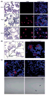
Representative images of monocyte/macrophage (MP) involvement in establishment of the PF-1 MCL cell line. (A) Hematoxylin and eosin (H&E) staining (left column), confocal microscopy analysis of CD68 + cells (red) (middle and right columns) of primary MCL cells in culture on days 0 (upper row), 7 (middle row) and 14 (bottom row). Topro3 was used to stain nuclei (blue). Original magnification, ×400. (B) H&E staining (left panel) and confocal microscopy analysis of CD68 + cells (right panel), showing the interaction between macrophages and MCL cells after 4 weeks of cell culturing. (C) No macrophage transformation in purified MCL cells. Primary cells were left unpurified (left column) or purified using the B-cell separation kit (right column). Cells were cultured for 14 days and then stained for CD68 (red) and Topro3 (blue, nuclear marker). Arrows in the light field indicate tumor cells undergoing apoptosis.
Figure 2
Figure 2
Morphologic and phenotypic features of PF-1 MCL cells. (A) Distribution of the size (longest diameter) of PF-1 MCL cells after 4 months of cell culturing. (B) Representative images of H&E-stained PF-1 MCL cells at different time points, 4 months (left panel) and 8 months (right panel) in tissue culture. (C) Representative immunophenotype flow cytometric histograms of PF-1 MCL cells. (Left) Immunostaining for both CD5 and CD19. (Right) Immunostaining for kappa and lambda light chains.
Figure 3
Figure 3
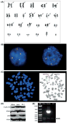
Conventional cytogenetics and FISH analysis of PF-1 MCL cells. (A) Representative karyotype of PF-1 MCL cells. Arrows point to abnormalities listed in Table II. (B) FISH detection of t(11;14)(q13;q32) in PF-1 MCL cells. IGH/CCND1 dual-color, dual-fusion translocation probes were used. Green: probe for the immunoglobulin heavy chain gene (14q32.2); red: probe for the CCDN1 gene (11q13); yellow: fusion gene signal. (C) Representative MapBack of FISH analysis showing t(11;14). (D) Western blot analysis of cyclin D1, CDK4 and p53 protein expressions in Mino and Jeko (two typical MCL cell lines) and PF-1 (new cell line) MCL cells. Actin served as an internal loading control. (E) EBV status in PF-1 cells. PCR analysis for EBNA2 gene in Mino (negative control), Granta (positive control) and PF-1 MCL cells. M, molecular marker; NS, non-specific band that served as an internal control.

References

    1. Dreyling M. Therapy of mantle cell lymphoma: new treatment options in an old disease or vice versa? Semin Hematol. 2011;48:145–147. - PubMed
    1. Jares P, Campo E. Advances in the understanding of mantle cell lymphoma. Br J Haematol. 2008;142:149–165. - PubMed
    1. Parekh S, Weniger MA, Wiestner A. New molecular targets in mantle cell lymphoma. Semin Cancer Biol. 2011;21:335–346. - PMC - PubMed
    1. Smith MR. Should there be a standard therapy for mantle cell lymphoma? Future Oncol. 2011;7:227–237. - PubMed
    1. Harris NL, Jaffe ES, Stein H, et al. A revised European-American classification of lymphoid neoplasms: a proposal from the International Lymphoma Study Group. Blood. 1994;84:1361–1392. - PubMed

Publication types

Substances

LinkOut - more resources