

Generation of β cells from human pluripotent stem cells: are we there yet?
- PMID: 24611778
- PMCID: PMC4144703
- DOI: 10.1111/nyas.12369
Generation of β cells from human pluripotent stem cells: are we there yet?
Abstract
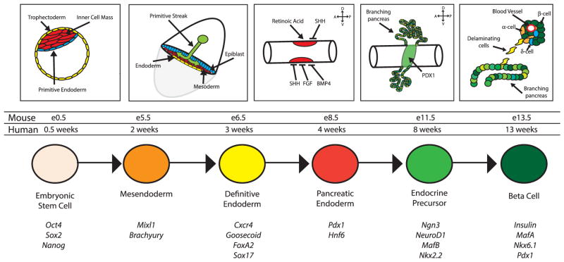
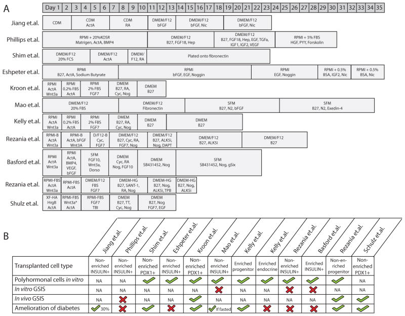
In 1998, the landmark paper describing the isolation and culture of human embryonic stem cells (ESCs) was published. Since that time, the main goal of many diabetes researchers has been to derive β cells from ESCs as a renewable cell-based therapy for the treatment of patients with diabetes. In working toward this goal, numerous protocols that attempt to recapitulate normal pancreatic development have been published that result in the formation of pancreatic cell types from human pluripotent cells. This review examines stem cell differentiation methods and places them within the context of pancreatic development. We additionally compare strategies that are currently being used to generate pancreatic cell types and contrast them with approaches that have been used to generate functional cell types in different lineages. In doing this, we aim to identify how new approaches might be used to improve yield and functionality of in vitro-derived pancreatic β cells as an eventual cell-based therapy for type 1 diabetes.
Keywords: diabetes; differentiation; endoderm; pancreas; pluripotent stem cell; β cell.
© 2014 New York Academy of Sciences.
Conflict of interest statement
The authors declare no conflicts of interest.
Figures


References
-
- Thomson JA, et al. Embryonic stem cell lines derived from human blastocysts. Science. 1998;282:1145–1147. - PubMed
-
- Reubinoff BE, et al. Embryonic stem cell lines from human blastocysts: somatic differentiation in vitro. Nat Biotechnol. 2000;18:399–404. - PubMed
-
- Takahashi K, et al. Induction of pluripotent stem cells from adult human fibroblasts by defined factors. Cell. 2007;131:861–872. - PubMed
-
- Tada S, et al. Characterization of mesendoderm: a diverging point of the definitive endoderm and mesoderm in embryonic stem cell differentiation culture. Development. 2005;132:4363–4374. - PubMed
-
- Lawson KA, Pedersen RA. Cell fate, morphogenetic movement and population kinetics of embryonic endoderm at the time of germ layer formation in the mouse. Development. 1987;101:627–652. - PubMed
Publication types
MeSH terms
Grants and funding
LinkOut - more resources
Full Text Sources
Other Literature Sources
