Editorial
. 2014 Aug;16(8):1133-45.
doi: 10.1111/cmi.12286. Epub 2014 Apr 3.

Anaplasma phagocytophilum surface protein AipA mediates invasion of mammalian host cells

Affiliations
Editorial

Anaplasma phagocytophilum surface protein AipA mediates invasion of mammalian host cells

David Seidman et al. Cell Microbiol. 2014 Aug.

Abstract

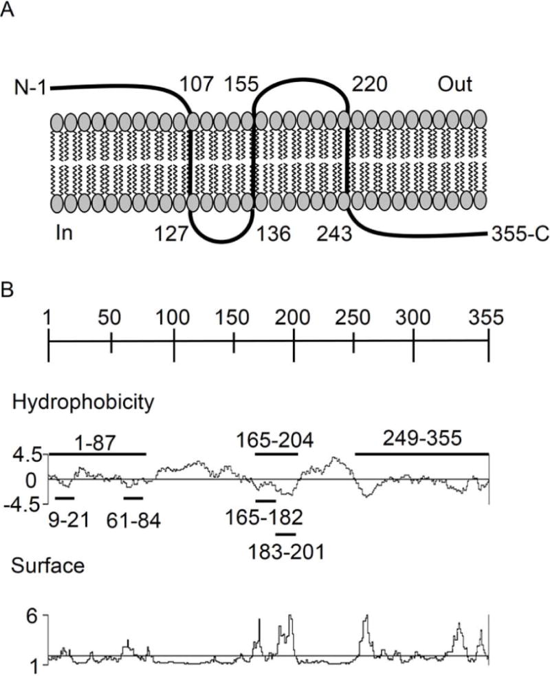
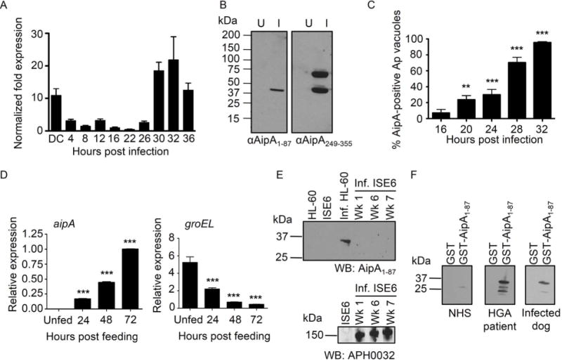
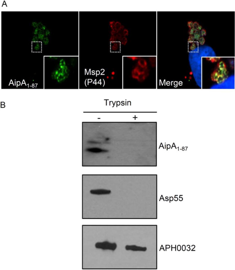
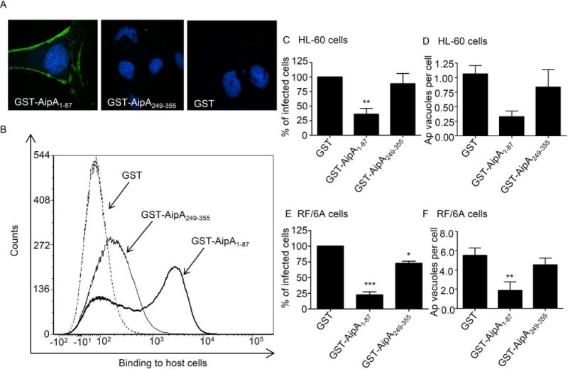
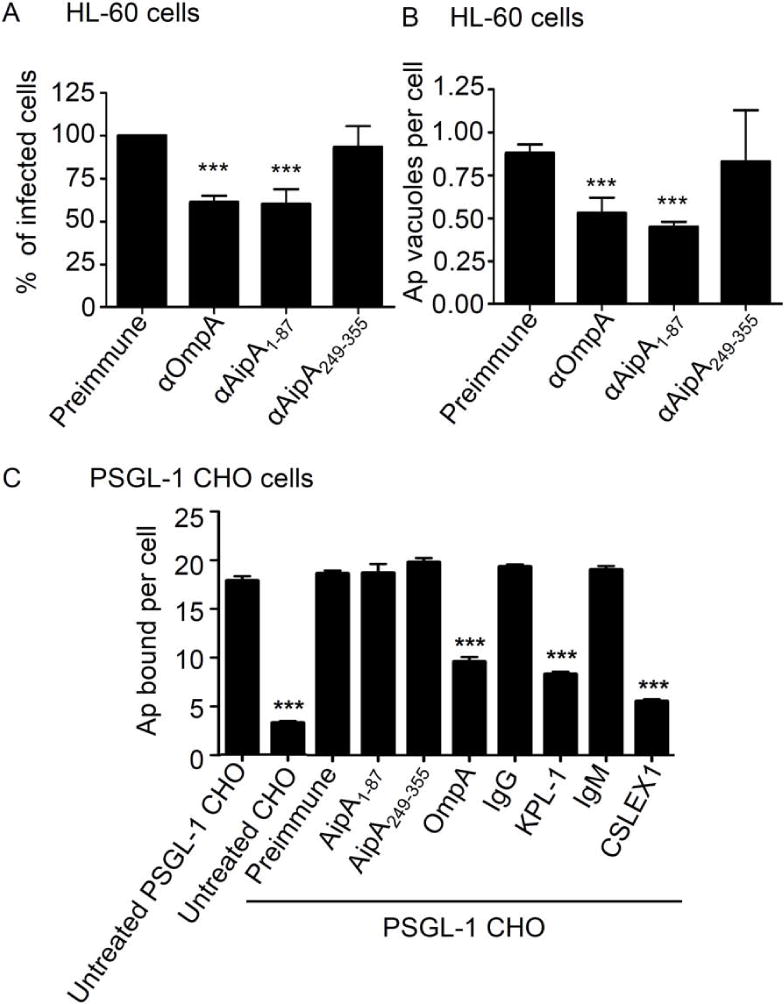
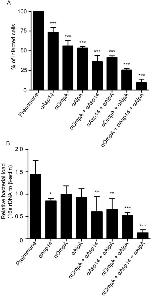
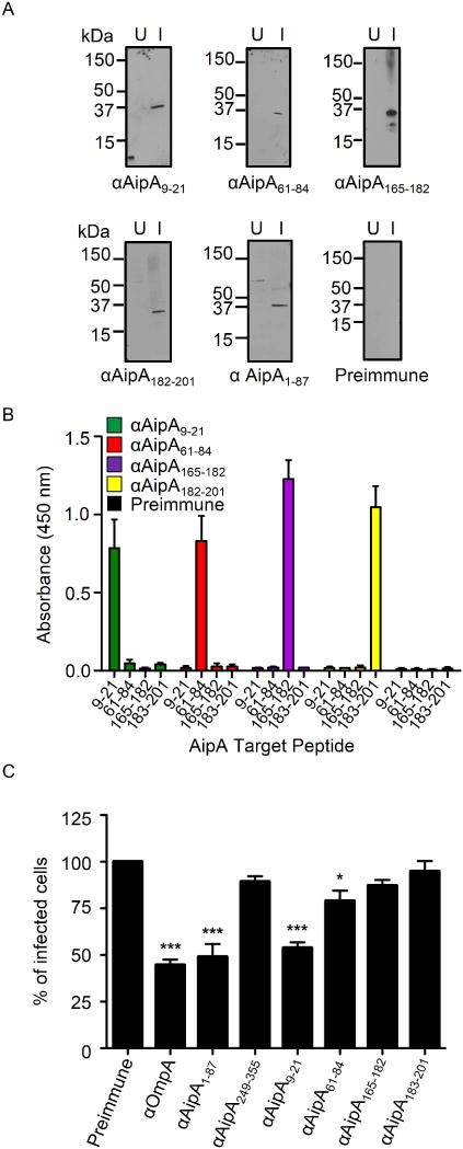
Anaplasma phagocytophilum, which causes granulocytic anaplasmosis in humans and animals, is a tick-transmitted obligate intracellular bacterium that mediates its own uptake into neutrophils and non-phagocytic cells. Invasins of obligate intracellular pathogens are attractive targets for protecting against or curing infection because blocking the internalization step prevents survival of these organisms. The complement of A. phagocytophilum invasins is incompletely defined. Here, we report the significance of a novel A. phagocytophilum invasion protein, AipA. A. phagocytophilum induced aipA expression during transmission feeding of infected ticks on mice. The bacterium upregulated aipA transcription when it transitioned from its non-infectious reticulate cell morphotype to its infectious dense-cored morphotype during infection of HL-60 cells. AipA localized to the bacterial surface and was expressed during in vivo infection. Of the AipA regions predicted to be surface-exposed, only residues 1 to 87 (AipA1-87 ) were found to be essential for host cell invasion. Recombinant AipA1-87 protein bound to and competitively inhibited A. phagocytophilum infection of mammalian cells. Antiserum specific for AipA1-87 , but not other AipA regions, antagonized infection. Additional blocking experiments using peptide-specific antisera narrowed down the AipA invasion domain to residues 9 to 21. An antisera combination targeting AipA1-87 together with two other A. phagocytophilum invasins, OmpA and Asp14, nearly abolished infection of host cells. This study identifies AipA as an A. phagocytophilum surface protein that is critical for infection, demarcates its invasion domain, and establishes a rationale for targeting multiple invasins to protect against granulocytic anaplasmosis.


Figures

Figure 1
Figure 1. Schematic diagrams of A. phagocytophilum AipA membrane topology and sequence
(A) Diagram of the predicted topology of AipA in the A. phagocytophilum outer membrane. N, AipA amino terminus. C, AipA carboxy terminus. Numerical values indicate amino acid coordinates for predicted transmembrane spanning regions. (B) Diagrams of the AipA sequence. The scale indicates 50-amino acid intervals. For the Hydrophobicity diagram, the Kyte-Doolitle algorithm was used to determine hydrophobic (histogram above the axis) and hydrophilic (histogram below axis) regions. For the Surface diagram, the Emini algorithm was used to determine regions that are likely accessible on the surface of the AipA (histogram above the axis) or not (histogram below the axis). The AipA amino acid segments against which antisera were raised are indicated on the Hydrophobicity plot by horizontal lines.
Figure 2
Figure 2. Differential expression profiling of AipA throughout the A. phagocytophilum life cycle
(A) aipA transcriptional profile during A. phagocytophilum infection of HL-60 cells. DC bacteria were incubated with HL-60 cells to establish a synchronous infection. Total RNA isolated from the DC inoculum and infected host cells at several postinfection time points was subjected to reverse transcriptase-quantitative PCR (RT-qPCR). Relative aipA transcript levels were normalized to A. phagocytophilum 16s rRNA gene transcript levels. To determine relative aipA transcription between RC and DC organisms, normalized aipA transcript levels per time point were calculated as the fold change in expression relative to expression at 16 h, a time point at which the entire bacterial population is in the DC morphotype. Data are the means ± standard deviations (SD) for triplicate samples and are representative of two experiments having similar results. (B) Western blot screening of whole-cell lysates of uninfected (U) and A. phagocytophilum infected HL-60 cells (I) using mouse antiserum raised against GST-AipA1–87 (αAipA1–87) and GST-AipA249–355 (αAipA249–355). (C) AipA expression over the course of infection of mammalian host cells. RF/6A cells that had been synchronously infected with A. phagocytophilum (Ap) were screened with antibodies targeting Msp2 (P44) (to denote all A. phagocytophilum inclusions) and AipA viewed by confocal microscopy. Data presented are the mean percentages ± SD of Msp2 (P44)-positive A. phagocytophilum inclusions that were also AipA-positive. At least 100 bacterial inclusions were scored per time point. (D) aipA and groEL expression during transmission feeding of A. phagocytophilum infected ticks on naïve mice. A. phagocytophilum-infected I. scapularis nymphs were allowed to feed on mice for 72 h. Total RNA recovered from unfed and transmission-fed ticks that had been removed at 24, 48, and 72 h postattachment was subjected to RT-qPCR. Relative aipA and groEL transcript levels were normalized to A. phagocytophilum 16S rRNA gene levels. (E) AipA is not expressed during A. phagocytophilum infection of a tick cell line. Western blot analysis of uninfected and A. phagocytophilum infected (Inf.) HL-60 and ISE6 cells using antiserum specific for AipA1–87 or APH0032. The number of weeks (Wk.) during which A. phagocytophilum was maintained in ISE6 cells are indicated. (F) AipA is expressed in vivo and elicits a humoral immune response. Western blot analysis of GST and GST-AipA1–87 screened with sera from an HGA patient and from an A. phagocytophilum infected dog. Results presented in panels B to F are each representative of at two to three independent experiments with similar results. Statistically significant (*, P < 0.05; **, P < 0.005; ***, P < 0.001) values are indicated.
Figure 3
Figure 3. AipA is an A. phagocytophilum OMP
(A) AipA colocalizes with the confirmed OMP, Msp2 (P44). A. phagocytophilum-infected RF/6A cells were fixed and viewed by confocal microscopy to assess immunoreactivity with AipA antiserum (green) in conjunction with Msp2 (P44) antiserum (red). Host cell nuclei were stained with DAPI (4′,6′-diamidino-2-phenylindole; blue). The insets demarcated by solid boxes in the lower right corners of each panel are magnified versions of the representative A. phagocytophilum-occupied vacuole that is denoted by the hatched box in each panel. (B) AipA is exposed on the bacterial surface. Intact A. phagocytophilum DC organisms were incubated with trypsin or vehicle control, solubilized, and Western-blotted. Immunoblots were screened with antiserum targeting AipA1–87, Asp55, or APH0032. Data are representative of two experiments with similar results.
Figure 4
Figure 4. GST-AipA requires amino acids 1 to 87 to bind and competitively inhibit A. phagocytophilum infection of mammalian host cells
(A and B) GST-AipA1–87 binds to mammalian host cells. RF/6A cells were incubated with GST-AipA1–87, GST-AipA249–355, or GST alone. (A) The host cells were fixed, screened with GST antibody (green), and examined using confocal microscopy. Host cell nuclei were stained with DAPI. Representative merged fluorescent images from three experiments with similar results are shown. (B) Flow cytometric analysis of GST fusion protein binding to RF/6A cells. (C to F) GST-AipA1–87 competitively inhibits A. phagocytophilum infection. HL-60 (C and D) and RF/6A cells (E and F) were incubated with DC bacteria in the presence of GST, GST-AipA1–87, or GST-AipA249–355 for 1 h. Following removal of unbound bacteria, host cells were incubated for 24 h (C and D) or 48 h (E and F) and subsequently examined using confocal microscopy to assess the percentage of infected cells (C and E) or the mean number (± SD) of pathogen-occupied vacuoles per cell (D and F). Results shown are relative to GST-treated host cells and are the means ± SD for three experiments. Statistically significant (*, P < 0.05; **, P < 0.005; ***, P < 0.001) values are indicated.
Figure 5
Figure 5. Pretreatment of A. phagocytophilum with AipA1–87 antiserum inhibits infection of HL-60 cells but does not alter binding to sLex-capped PSGL-1
A. phagocytophilum DC organisms were exposed to antiserum targeting AipA1–87, AipA249–355, OmpA, or preimmune serum and then incubated with HL-60 (A and B), PSGL-1 CHO cells, or untransfected CHO cells (C). The infection of HL-60 cells was allowed to proceed for 24 h prior to being assessed, while bacterial binding to PSGL-1 CHO cells was assessed immediately. The mean ± standard deviations of percentages of infected HL-60 cells (A), A. phagocytophilum (Ap) vacuolar inclusions per HL-60 cell (B), and bound DC organisms per PSGL-1 CHO cell or untransfected CHO cell (C) were determined using immunofluorescence microscopy. Additional positive controls for blocking A. phagocytophilum to PSGL-1 CHO cells, besides incubating bacteria with OmpA antiserum, were PSGL-1 CHO cells that had been incubated with PSGL-1 N-terminus blocking antibody KPL-1 or sLex-blocking antibody CSLEX1 prior to the addition of bacteria. Negative controls were PGSL-1 CHO cells that had been incubated with isotype control antibodies prior to the addition of bacteria. Results shown are relative to GST-treated host cells and are the means ± SD for three experiments. Statistically significant (***, P < 0.001) values are indicated.
Figure 6
Figure 6. A combination of antisera targeting AipA, OmpA, and Asp14 blocks A. phagocytophilum infection of host cells
DC bacteria were incubated with preimmune serum or antiserum targeting AipA1–87, OmpA, and/or Asp14 and then incubated with HL-60 cells. (A) The cells were fixed and screened by confocal microscopy to assess the percentage of infected cells. Results shown are relative to host cells that had been treated with preimmune serum and are the means ± SD for three experiments. (B) DNA isolated from the cells was subjected to quantitative PCR analyses. Relative DNA loads of A. phagocytophilum 16s rRNA gene were normalized to DNA loads of the human β-actin gene. Results shown are the means ± SD of triplicate samples and are representative of three independent experiments with similar results. Statistically significant (*, P < 0.05; **, P < 0.005; ***, P < 0.001) values relative to the bacterial load of host cells that had been incubated with preimmune antisera are presented.
Figure 7
Figure 7. AipA residues 9–21 are critical for establishing infection in host cells
(A) Western blot analyses in which rabbit antiserum targeting AipA9–21, AipA61–84, AipA165–182, AipA183–201, AipA1–87, or preimmune rabbit serum was used to screen whole cell lysates of uninfected (U) and A. phagocytophilum infected HL-60 cells (I). Data are representative of two experiments with similar results. (B) ELISA in which AipA9–21, AipA61–84, AipA165–182, and AipA183–201 antibodies were used to screen wells coated with peptides corresponding to AipA residues 9–21, 61–84, 165–182 and 183–201. Each antiserum only recognized the peptide against which it had been raised. Results shown are the mean (± SD) of triplicate samples. Data are representative of three experiments with similar results. (C) Pretreatment of A. phagocytophilum with AipA9–21 antiserum inhibits infection of HL-60 cells. DC bacteria were pretreated with antiserum specific for AipA9–21, AipA61–84, AipA165–182, AipA183–201, AipA1–87, AipA249–355, OmpA, or preimmune serum for 30 min. Next, the treated bacteria were incubated with HL-60 cells for 60 min. After removal of unbound bacteria, host cells were incubated for 24 h and subsequently examined using Msp2 (P44) antibody and confocal microscopy to assess the percentage of infected cells. Results shown are relative to preimmune serum-treated host cells and are the means ± SD for six experiments. Statistically significant (*, P < 0.05; **, P < 0.005; ***, P < 0.001) values are indicated.

References

    1. Alhumaidan H, Westley B, Esteva C, Berardi V, Young C, Sweeney J. Transfusion-transmitted anaplasmosis from leukoreduced red blood cells. Transfusion. 2013;53:181–186. - PubMed
    1. Annen K, Friedman K, Eshoa C, Horowitz M, Gottschall J, Straus T. Two cases of transfusion-transmitted Anaplasma phagocytophilum. American journal of clinical pathology. 2012;137:562–565. - PubMed
    1. Barbosa AS, Abreu PA, Neves FO, Atzingen MV, Watanabe MM, Vieira ML, et al. A newly identified leptospiral adhesin mediates attachment to laminin. Infection and immunity. 2006;74:6356–6364. - PMC - PubMed
    1. Brennan MJ, Shahin RD. Pertussis antigens that abrogate bacterial adherence and elicit immunity. American journal of respiratory and critical care medicine. 1996;154:S145–149. - PubMed
    1. Cardwell MM, Martinez JJ. The Sca2 autotransporter protein from Rickettsia conorii is sufficient to mediate adherence to and invasion of cultured mammalian cells. Infection and immunity. 2009;77:5272–5280. - PMC - PubMed

Publication types

MeSH terms