. 2014 Mar 10;5(1):8.
doi: 10.1186/1759-8753-5-8.

Bacterial group I introns: mobile RNA catalysts

Affiliations

Bacterial group I introns: mobile RNA catalysts

Georg Hausner et al. Mob DNA. .

Abstract

Group I introns are intervening sequences that have invaded tRNA, rRNA and protein coding genes in bacteria and their phages. The ability of group I introns to self-splice from their host transcripts, by acting as ribozymes, potentially renders their insertion into genes phenotypically neutral. Some group I introns are mobile genetic elements due to encoded homing endonuclease genes that function in DNA-based mobility pathways to promote spread to intronless alleles. Group I introns have a limited distribution among bacteria and the current assumption is that they are benign selfish elements, although some introns and homing endonucleases are a source of genetic novelty as they have been co-opted by host genomes to provide regulatory functions. Questions regarding the origin and maintenance of group I introns among the bacteria and phages are also addressed.


Figures

Figure 1
Figure 1
The distribution and diversity of group I introns. A small subunit rDNA cladogram shows the biological host range for each group I intron subclass in bacteria (B) and viruses (V). Distribution of group I introns in Eukarya as well as the cellular location of each subclass is indicated (N, nucleus; M, mitochondria; C, chloroplast). This figure was generated based on the available information obtained from the Comparative RNA Website [http://www.rna.icmb.utexas.edu/] and Group I Intron Sequence and Structure Database [http://www.rna.whu.edu.cn/gissd/index.html].
Figure 2
Figure 2
Secondary structure model for group I introns. Generic secondary structure representations for group I introns highlighting the locations of intron-encoded proteins. (a) The blue lines indicate regions where ORFs that encode homing endonucleases are entirely located in loops. (b) In some group I introns, the endonuclease ORFs extend and overlap with intron core sequences. In both panels, stem regions are represented by solid black lines and single-stranded loop regions are represented by grey curved lines. Exon sequences are represented by black boxes. The ten pairing regions (P1 to P10) are also indicated. The solid green arrowheads indicate the intron-exon junctions (5′ and 3′ splicing sites). The positions of the internal guide sequence (IGS) and the so called P, Q, R and S sequence elements are indicated by thick orange lines. The guanosine-5’-triphosphate (GTP) binding pocket within the P7 helix is indicated by an asterisk.
Figure 3
Figure 3
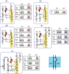
Differences between group I intron classes (IA to IE). Shown are secondary structure representatives for the group I intron classes [26-33]. The IA to ID classes are commonly found in bacteria. The IE class is also depicted for comparative purposes. For all group I intron RNA structures the catalytic core is highlighted in yellow. Beside each secondary structure model is a sequence logo alignment of the P7:P7′ pairing for the intron subclasses. The P7:P7′ pairing is important because it is a highly conserved region and is diagnostic for discriminating between various group I intron subclasses. With regards to the sequence logos the information content at each position (in bits, from 0 to 2) is represented by the height of the nucleotide. A score of 2 bits corresponds to high conservation, while a score of 0 corresponds to low conservation. The number of sequences used to generate each sequence logo is indicated below the intron subtype. Asterisks indicate the possible locations of peripheral insertions within the intron. The catalytic domain is highlighted in yellow.
Figure 4
Figure 4
Schematic representation of group I intron splicing. The splicing pathway consists of two sequential transesterification reactions. The first reaction is initiated by the 3′–OH group of an exogenous GTP (αG) that docks into the G-binding pocket located in the P7 region and the 3′–OH group attacks the 5′ splice site. In the second reaction, the 3′–OH of the released 5′ exon attacks the phosphodiester bond between the intronic terminal G (ωG) and the 3′ exon, resulting in the liberation of the intron and the ligation of the exons.
Figure 5
Figure 5
Mobility pathways mediated by homing endonucleases. Schematics of different endonuclease-mediated mobility pathways between donor and recipient alleles. (a) group I intron homing mediated by intron-encoded endonucleases; (b) the collaborative or trans homing pathway; (c) the intronless homing pathway mediated by free-standing endonucleases. In all cases, the homing endonuclease gene is represented by a green rectangle, and the homing site of the endonuclease is shown by a grey filled rectangle. The green rectangle outlined with dashed line indicates the outcome of a recombination event whereby the endonuclease ORF becomes embedded within an endonuclease-lacking intron, creating a potential mobile group I intron.

References

    1. Kruger K, Grabowski PJ, Zaug AJ, Sands J, Gottschling DE, Cech TR. Self-splicing RNA: autoexcision and autocyclization of the ribosomal RNA intervening sequence of Tetrahymena. Cell. 1982;31:147–157. doi: 10.1016/0092-8674(82)90414-7. - DOI - PubMed
    1. Michel F, Jacquier A, Dujon B. Comparison of fungal mitochondrial introns reveals extensive homologies in RNA secondary structure. Biochimie. 1982;64:867–881. doi: 10.1016/S0300-9084(82)80349-0. - DOI - PubMed
    1. Cech TR. Self-splicing of group-I introns. Annu Rev Biochem. 1990;55:599–629. - PubMed
    1. Lang BF, Laforest MJ, Burger G. Mitochondrial introns: a critical view. Trends Genet. 2007;23:119–125. doi: 10.1016/j.tig.2007.01.006. - DOI - PubMed
    1. Dujon B. Group I introns as mobile genetic elements: facts and mechanistic speculation - a review. Gene. 1989;82:91–114. doi: 10.1016/0378-1119(89)90034-6. - DOI - PubMed