Review
. 2014 May:17:74-80.
doi: 10.1016/j.dnarep.2014.02.006. Epub 2014 Mar 7.

Non-homologous end joining often uses microhomology: implications for alternative end joining

Affiliations
Review

Non-homologous end joining often uses microhomology: implications for alternative end joining

Nicholas R Pannunzio et al. DNA Repair (Amst). 2014 May.

Abstract

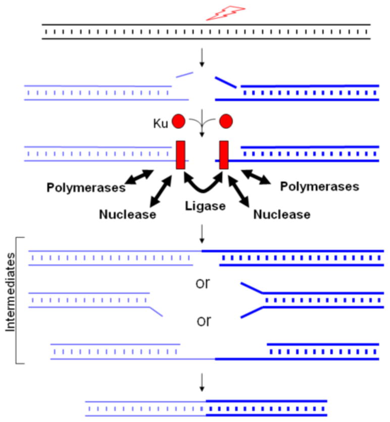
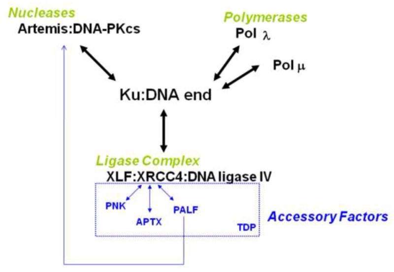
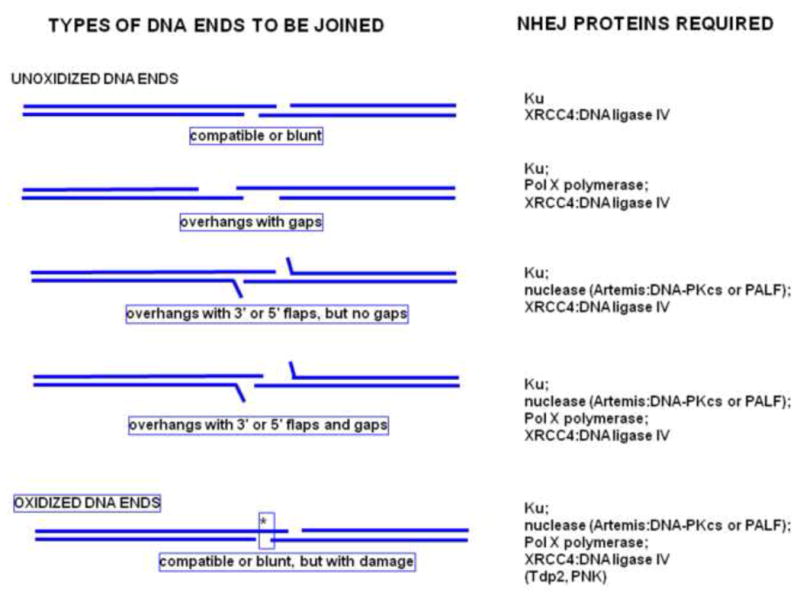
Artemis and PALF (also called APLF) appear to be among the primary nucleases involved in non-homologous end joining (NHEJ) and responsible for most nucleolytic end processing in NHEJ. About 60% of NHEJ events show an alignment of the DNA ends that use 1 or 2bp of microhomology (MH) between the two DNA termini. Thus, MH is a common feature of NHEJ. For most naturally occurring human chromosomal deletions (e.g., after oxidative damage or radiation) and translocations, such as those seen in human neoplasms and as well as inherited chromosomal structural variations, MH usage occurs at a frequency that is typical of NHEJ, and does not suggest major involvement of alternative pathways that require more extensive MH. Though we mainly focus on human NHEJ at double-strand breaks, comparison on these points to other eukaryotes, primarily S. cerevisiae, is informative.

Keywords: Double-strand break repair, Lymphoma, Chromosomal rearrangements, V(D)J recombination, Class switch recombination.


Figures

Figure 1
Figure 1. General steps of NHEJ
The lightning arrow indicates ionizing radiation (IR), a reactive oxygen species (ROS), or an enzymatic cause of a DSB. Ku binding to the DNA ends at double-strand breaks (DSBs) improves binding by nuclease, DNA polymerase, and ligase components. Note that Ku is thought to change conformation upon binding to the DNA end, as depicted by its shape change from a sphere to a rectangle. Flexibility in the loading of these enzymatic components, the option to load repeatedly (iteratively), and independent processing of the two DNA ends all permit mechanistic flexibility for the NHEJ process. This mechanistic flexibility is essential to permit NHEJ to handle a very diverse array of DSB end configurations and to join them. In addition to the overall mechanistic flexibility, each component exhibits enzymatic flexibility and multifunctionality. The figure shows that there are many alternative intermediates in the joining process. These intermediates are reflected in a diverse DNA sequences at the junction of the joining process.
Figure 2
Figure 2. Proteins Involved in Vertebrate NHEJ
The diagram shows known interactions of the proteins documented to participate in the key enzymatic steps of vertebrate NHEJ. (Other proteins may participate in gaining access to the chromatinized DNA or in modulating some of the components shown [70, 71].) XLF, also called Cernunnos, is a component of the DNA ligase IV complex [72, 73]. PNK is polynucleotide kinase. APTX is aprataxin [74]. PALF (Polynucleotide kinase and Aprataxin-like Fork-head-associated) and is more commonly called APLF, based on an alternative order of the same acronym; and TDP is tyrosyl DNA phosphodiesterase (see article in this series by K. W. Caldecott).
Figure 3
Figure 3. DNA End Configurations Determine Which NHEJ Proteins are Required During NHEJ
The distinctive flexibility required by NHEJ means that it is not easily depicted as a simple linear pathway. If NHEJ were simple enough to depict in a linear pathway, then it would probably not be sufficiently flexible to handle many different DNA end configurations, only a subset of which are illustrated here. Not illustrated is the likely aspect that NHEJ appears to be iterative. The nucleases, polymerases, and the ligase complex can work on either of the two DNA ends independently, iteratively, and in any order until both strands are ligated. Some possible end configurations and corresponding required proteins are listed. The asterisk in the bottom joining diagram represents oxidative base damage.
Figure 4
Figure 4. Two models of NHEJ
A. In one conceptualization, two separate NHEJ pathways exist, classical NHEJ (c-NHEJ) and alternative NHEJ (alt-NHEJ) with the latter occurring independently of c-NHEJ components and relying on increased lengths of microhomology (MH) and for a higher percentage of the junctions. B. In a conceptualization that we favor, there is only one NHEJ pathway with alternative components able to substitute for some of the canonical NHEJ components. In WT cells, the alternative components are infrequently used because the c-NHEJ components are most efficient. But if a cell is deficient in a major NHEJ factor, it must utilize the alternative components to complete the join (e.g., L1 or L3 in place of L4), but perhaps with reduced kinetic efficiency. Note that when an NHEJ component is missing, incomplete intermediates of joining may accumulate, as indicated by the thicker blue line.

References

    1. Lieber MR. The Mechanism of Double-Strand DNA Break Repair by the Nonhomologous DNA End-Joining Pathway. Annu Rev Biochem. 2010;79:181–211. - PMC - PubMed
    1. Ma Y, Lu H, Schwarz K, Lieber MR. Repair of double-strand DNA breaks by the human nonhomologous DNA end joining pathway: the iterative processing model. Cell Cycle. 2005;4:1193–1200. - PubMed
    1. Moshous D, Li L, Chasseval R, Philippe N, Jabado N, Cowan MJ, Fischer A, Villartay J-Pd. A new gene involved in DNA double-strand break repair and V(D)J recombination is located on human chromosome 10p. Hum Mol Gen. 2000;9:583–588. - PubMed
    1. Li L, Moshous D, Zhou Y, Wang J, Xie G, Salido E, Hu D, Villartay JPd, Cowan MJ. A founder mutation in Artemis, an SNM1-like protein, causes SCID in Athabascan-speaking Native Americans. J Immunol. 2002;168:6323–6329. - PubMed
    1. Rooney S, Alt FW, Lombard D, Whitlow S, Eckersdorff M, Fleming J, Fugmann S, Ferguson DO, Schatz DG, Sekiguchi J. Defective DNA repair and increased genomic instability in Artemis-deficient murine cells. J Exp Med. 2003;197:553–565. - PMC - PubMed

Substances