. 2014 Mar 10;9(3):e90913.
doi: 10.1371/journal.pone.0090913. eCollection 2014.

Extracellular vesicles in luminal fluid of the ovine uterus

Affiliations

Extracellular vesicles in luminal fluid of the ovine uterus

Gregory Burns et al. PLoS One. .

Abstract

Microvesicles and exosomes are nanoparticles released from cells and can contain small RNAs, mRNA and proteins that affect cells at distant sites. In sheep, endogenous beta retroviruses (enJSRVs) are expressed in the endometrial epithelia of the uterus and can be transferred to the conceptus trophectoderm. One potential mechanism of enJSRVs transfer from the uterus to the conceptus is via exosomes/microvesicles. Therefore, studies were conducted to evaluate exosomes in the uterine luminal fluid (ULF) of sheep. Exosomes/microvesicles (hereafter referred to as extracellular vesicles) were isolated from the ULF of day 14 cyclic and pregnant ewes using ExoQuick-TC. Transmission electron microscopy and nanoparticle tracking analysis found the isolates contained vesicles that ranged from 50 to 200 nm in diameter. The isolated extracellular vesicles were positive for two common markers of exosomes (CD63 and HSP70) by Western blot analysis. Proteins in the extracellular vesicles were determined by mass spectrometry and Western blot analysis. Extracellular vesicle RNA was analyzed for small RNAs by sequencing and enJSRVs RNA by RT-PCR. The ULF extracellular vesicles contained a large number of small RNAs and miRNAs including 81 conserved mature miRNAs. Cyclic and pregnant ULF extracellular vesicles contained enJSRVs env and gag RNAs that could be delivered to heterologous cells in vitro. These studies support the hypothesis that ULF extracellular vesicles can deliver enJSRVs RNA to the conceptus, which is important as enJSRVs regulate conceptus trophectoderm development. Importantly, these studies support the idea that extracellular vesicles containing select miRNAs, RNAs and proteins are present in the ULF and likely have a biological role in conceptus-endometrial interactions important for the establishment and maintenance of pregnancy.


Conflict of interest statement

Competing Interests: The authors have declared that no competing interests exist.

Figures

Figure 1
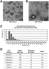
Figure 1. Transmission electron microscopy (TEM) and nanoparticle tracking analysis (NTA) of vesicles isolated from uterine luminal fluid of sheep.
(A–B) TEM revealed vesicles of about 100 nm, consistent with exosomes. (C) NTA profile of extracellular vesicle collection from one individual. Measurements were collected from 3 samples; error bars represent +/−1 standard deviation. The profile confirms size estimates from TEM images. (D) Nanoparticle Tracking Analysis of collected extracellular vesicles. Precipitated extracellular vesicles were suspended in PBS+0.1%BSA and analyzed by NTA using a NanoSight instrument. Mean diameter, standard deviation of the mean, and mode size are reported (Replicates ≥3 per sample). These measurements are consistent with the presence of exosomes and microvesicles in the collected samples from ULF.
Figure 2
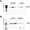
Figure 2. Western blot showing presence of HSP70 and CD63 in ULF derived extracellular vesicles from cyclic and pregnant ewes.
Western blot analysis shows immunoreactive bands for (A) HSP70 and (B), CD63 two well characterized exosome markers, in all samples.
Figure 3
Figure 3. Proteomic analysis of cyclic and pregnant ULF extracellular vesicles.
(A) Venn diagram containing 195 unique proteins identified by nano-LC-MS/MS analysis with 40 and 76 unique to the 14C and 14P groups respectively and 79 proteins in common. Note the presence of CTSL1 in both samples along with the presence of PTGS2 in only the 14P sample. (B) Western blot analysis shows the presence of CTSL in all samples. (C) PTGS2 immunoreactive bands are absent in 14C samples and present in 14P (2/3) samples.
Figure 4
Figure 4. RNA content of extracellular vesicles.
(A) The RNA profiles of extracellular vesicles, endometrium and conceptus samples using an Agilent 2100 Bioanalyzer. Note the cluster of RNA species at or below 200 base pairs in the extracellular vesicle samples both cyclic and pregnant; in contrast to endometrium and conceptus samples which display profiles characteristic of eukaryotic tissue with clearly defined 18S and 28S rRNA peaks and comparatively low percentages of transcripts below 200 base pairs. (B) Endogenous Jaagsiekte retrovirus (enJRSVs) env and gag RNA is present in extracellular vesicles from ULF of cyclic and pregnant ewes by RT-PCR. Day 14 conceptus and endometrium were used as positive controls for env. RT-PCR for gag utilized RNA from day 14 conceptus and 100 day placentome as positive controls. Negative control (NC) was performed by omitting cDNA in the reaction mix.
Figure 5
Figure 5. Small RNA sequencing of ULF derived extracellular vesicles.
Next generation small RNA sequencing analysis restricted to at least 10 copies of each sequence from 15–55 nucleotides in length and grouped by mature miRNA produced 81 mature bovine miRNA species referenced in miRBase (Release 19). A total of 53 mature miRNAs were common to cyclic and pregnant samples with one unique to the 14P group and 27 unique to the 14C group.
Figure 6
Figure 6. Cellular and extracellular detection of mature miRNA by RT-qPCR in cyclic and pregnant endometrium, conceptus and ULF extracellular vesicles.
Presence of selected mature miRNAs was tested for in cyclic and pregnant endometrium, conceptus, and extracellular vesicles from cyclic and pregnant ewes using the Qiagen miScript system and mature miRNA primer assays (400 pg cDNA/well). Expression values (2−ΔΔCt) were calculated using the geometric mean of SNORD95 and SNORD96A references and the geometric mean of the target gene. Error bars represent ± 1 SEM.
Figure 7
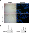
Figure 7. Delivery of enJSRVs env and gag RNA to HEK-293 cells.
(A) Brightfield and epifluorescent images of HEK-293 cells, counterstained with DAPI, untreated or exposed to extracellular vesicles labeled with PKH67 green fluorescent linker dye. (B) RT-PCR analysis of cell lysate for enJSRVs env and gag 48 hours after treatment with labeled extracellular vesicles.

References

    1. Spencer TE, Sandra O, Wolf E (2008) Genes involved in conceptus-endometrial interactions in ruminants: insights from reductionism and thoughts on holistic approaches. Reproduction 135: 165–179. - PubMed
    1. Spencer TE, Johnson GA, Bazer FW, Burghardt RC, Palmarini M (2007) Pregnancy recognition and conceptus implantation in domestic ruminants: roles of progesterone, interferons and endogenous retroviruses. Reprod Fertil Dev 19: 65. - PubMed
    1. Spencer TE, Bazer FW (2004) Conceptus signals for establishment and maintenance of pregnancy. Reprod Biol Endocrinol 2: 49. - PMC - PubMed
    1. Guillomot M (1995) Cellular interactions during implantation in domestic ruminants. J Reprod Fertil Suppl 49: 39–51. - PubMed
    1. Fléchon JE, Guillomot M, Charlier M, Fléchon B, Martal J (1986) Experimental studies on the elongation of the ewe blastocyst. Reprod Nutr Dev 26: 1017–1024. - PubMed

Publication types