. 2014 Apr;75(4):469-86.
doi: 10.1002/ana.24132. Epub 2014 Apr 14.

Brain injury in premature neonates: A primary cerebral dysmaturation disorder?

Affiliations

Brain injury in premature neonates: A primary cerebral dysmaturation disorder?

Stephen A Back et al. Ann Neurol. 2014 Apr.

Abstract

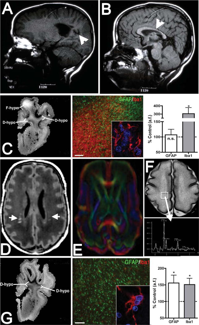
With advances in neonatal care, preterm neonates are surviving with an evolving constellation of motor and cognitive disabilities that appear to be related to widespread cellular maturational disturbances that target cerebral gray and white matter. Whereas preterm infants were previously at high risk for destructive brain lesions that resulted in cystic white matter injury and secondary cortical and subcortical gray matter degeneration, contemporary cohorts of preterm survivors commonly display less severe injury that does not appear to involve pronounced glial or neuronal loss. Nevertheless, these milder forms of injury are also associated with reduced cerebral growth. Recent human and experimental studies support that impaired cerebral growth is related to disparate responses in gray and white matter. Myelination disturbances in cerebral white matter are related to aberrant regeneration and repair responses to acute death of premyelinating late oligodendrocyte progenitors (preOLs). In response to preOL death, early oligodendrocyte progenitors rapidly proliferate and differentiate, but the regenerated preOLs fail to normally mature to myelinating cells required for white matter growth. Although immature neurons appear to be more resistant to cell death from hypoxia-ischemia than glia, they display widespread disturbances in maturation of their dendritic arbors, which further contribute to impaired cerebral growth. These complex and disparate responses of neurons and preOLs thus result in large numbers of cells that fail to fully mature during a critical window in development of neural circuitry. These recently recognized forms of cerebral gray and white matter dysmaturation raise new diagnostic challenges and suggest new therapeutic directions centered on reversal of the processes that promote dysmaturation.


Conflict of interest statement

Potential Conflicts of Interest

Nothing to report.

Figures

FIGURE 1
FIGURE 1
Microscopic necrosis has a high incidence but constitutes a small fraction of preterm cerebral white matter injury (WMI). (A) Typical sparse distribution of human microcysts visualized by staining for β-amyloid precursor protein, a marker of axonal degeneration. Note that axonal degeneration is usually restricted to microcysts and is not visualized in the surrounding regions of diffuse WMI. Sample is from a human autopsy brain at 32 weeks postconception. (B) Detail of the degenerating axons in the microcyst seen in the box in A. Degenerating axons were visualized both in the core and at the periphery of the microcyst. (C) Detail of the degenerating axons in the box in B shows numerous swollen and dystrophic-appearing axons. (D) A microcyst visualized by high-field (12T; T2) ex vivo magnetic resonance imaging as a focal hypointense lesion (F-hypo). Chronic WMI was analyzed 2 weeks after global cerebral ischemia in a fetal sheep model of preterm cerebral injury. Note that the surrounding diffuse WMI appeared as a diffuse hyperintense signal (D-hyper). (E) Pie chart showing the approximate relative percentages of human diffuse WMI, cystic periventricular leukomalacia (PVL), and microscopic necrosis, adapted from Pierson et al and Buser et al. (F) Schematic diagram showing the relative burden of human microcysts (dots) relative to diffuse WMI (within dotted lines), which typically comprises >80% of the total burden of WMI. Scale bars: A, 500μm; B, 100μm; C, 25μm. [Color figure can be viewed in the online issue, which is available at wileyonlinelibrary.com.]
FIGURE 2
FIGURE 2
Myelination failure in chronic diffuse white matter injury (WMI) coincides with lesions highly enriched in reactive astrocytes, activated microglia, and oligodendrocyte progenitors (preOLs) that are arrested in their maturation. (A) Distinctly different pathogenetic mechanisms mediate abnormal myelination in necrotic lesions (periventricular leukomalacia [PVL]; upper pathway) versus lesions with diffuse WMI (lower pathway). Hypoxia–ischemia (HI) is illustrated as one potential trigger for WMI. More severe HI triggers white matter necrosis (upper pathway) with pancellular degeneration that depletes the white matter of glia and axons. Severe necrosis results in cystic PVL, whereas milder necrosis results in microcysts. Milder HI (lower pathway) selectively triggers early preOL death. preOLs are rapidly regenerated from a pool of early preOLs that are resistant to HI. Chronic lesions are enriched in reactive glia (astrocytes and microglia/macrophages) generating inhibitory signals that block preOL differentiation to mature myelinating oligodendrocytes. Myelination failure in diffuse WMI thus results from preOL arrest rather than axonal degeneration. The molecular mechanisms that trigger preOL arrest are likely to be multifactorial and related to factors intrinsic and extrinsic to the preOLs. Note that the lower pathway is the dominant one in most contemporary preterm survivors, whereas the minor upper pathway reflects the declining burden of white matter necrosis that has accompanied advances in neonatal intensive care. (B) Typical appearance of normal early myelination of axons in a perinatal rodent at postnatal day 10. Axons are visualized by staining for neurofilament protein (NF; red). Early myelination of axons is visualized with the O4 antibody (green). (C) preOL arrest in a chronic white matter lesion where numerous preOLs (green) are seen, but the axons (red) are diffusely unmyelinated. Scale bars = 100μm.
FIGURE 3
FIGURE 3
In premyelinated white matter (WM), the cellular mechanisms and extent of neuroaxonal degeneration and myelination failure are distinct for necrotic and diffuse WM injury (WMI). WM necrosis (cystic periventricular leukomalacia/microcysts; left panel) is characterized by loss of all cellular elements in necrotic foci (glia, axons, and interstitial neurons). Degeneration of axons and oligodendrocytes (OLs) both contribute to myelination failure. Retrograde degeneration of axons in necrotic foci contributes to neuronal loss in cerebral gray matter. Diffuse WMI (right panel) involves selective degeneration of OL progenitors (preOLs) with sparing of premyelinating axons and interstitial neurons. Note that recent experimental data support a role for selective vulnerability of larger caliber early myelinating axons that appear later in white matter development as myelination progresses. Myelination failure is related to a failure of preOL differentiation (preOL arrest) to OLs. The mechanism of neuronal dysmaturation is unclear and may involve a direct effect of gray matter ischemia on maturation of dendrites and spines as well as axonal factors related to chronic white matter inflammation.
FIGURE 4
FIGURE 4
The preterm brain is enriched in immature neurons that do not degenerate in response to ischemia, but are highly susceptible to impaired maturation that manifests as a less mature dendritic arbor with reduced spine density. (A) A typical pyramidal neuron from the preterm cerebral cortex of a control fetal sheep. Note the paucity of processes in contrast to the highly complex dendritic arbor of a pyramidal neuron from a near-term animal (B). (C, D) In response to preterm ischemia, cortical pyramidal neurons display disrupted maturation. Note that the typical control cell (C) is more highly arborized in contrast to the response to transient cerebral ischemia that resulted in a more simplified dendritic arbor (D). The relative complexity of the cells can be appreciated from the overlay of the red concentric Scholl rings, which illustrates that the processes of the dysmature neurons intersect less frequently with the rings. The yellow, white, pink, green, and blue lines represent first-, second-, third-, fourth-, and fifth-order branches, respectively, from the soma. Note the overall reduction in the size and complexity of the branching pattern in D. (E) Reductions in cortical growth also manifest as disturbances in cortical anisotropy. Note the normal progressive decline in fractional anisotropy (FA) in controls (blue) between preterm and near-term cortical development, as adapted from Dean et al. In response to ischemia, higher cortical anisotropy (more restricted water diffusion) was observed in response to ischemia (red) relative to control (blue), which was related to the reduced complexity of the dendritic arbor of the ischemic neurons (eg, in D) versus controls (eg, in C). Scale bars = 20nm. HI = hypoxia–ischemia.
FIGURE 5
FIGURE 5
Diagnostic and experimental magnetic resonance imaging (MRI) approaches to define dysmaturation processes related to white matter injury (WMI). (A, B) MRI (1.5T; T1) appearance of marked cystic necrotic human WMI (A; arrowhead) adjacent to the lateral ventricle, which is associated with white matter volume loss that involves the corpus callosum (B; arrowhead). (C) High-field MRI–defined focal necrotic WMI and corresponding histopathological features. WMI was analyzed at 1 or 2 weeks after global cerebral ischemia in 0.65 gestation fetal sheep (adapted from Riddle et al). At left is a representative T2-weighted image of a large focal hyperintense (F-hyper) lesion detected at 1 week. Note the hypointense diffuse WMI (D-hypo), discussed in G, below. In the center is a typical necrotic lesion defined by focal staining for reactive microglia and macrophages with Iba1 (red and inset) and a paucity of glial fibrillary acidic protein (GFAP)-labeled astrocytes. Nuclei in the inset are visualized with Hoechst 33342 (blue). At right, by 2 weeks after ischemia, necrotic WMI displayed a progressive decrease in GFAP-labeled astrocytes and a pronounced increased in Iba1-labeled macrophages and microglia. *p < 0.05; bar in C = 100μm. n.s. = nonsignificant. (D) Diffuse human WMI on diagnostic MRI (1.5T; T1) has the appearance of bilateral multifocal signal hyperintensities (arrows). (E) Diffusion tensor imaging (DTI) defines the microstructure of white matter tracts and can be used to follow the chronic progression of diffuse WMI. (F) Magnetic resonance spectroscopic imaging (MRSI) can be applied to define biochemical and metabolic abnormalities associated with diffuse WMI. Both DTI and MRSI detect abnormalities beyond the areas of signal abnormality on T1-weighted images. (G) Diffuse fetal ovine WMI defined at 12T as in C. At left is a representative image of diffuse hypointense (D-hypo) WMI seen on a T2-weighted image at 1 week after ischemia. In the center are typical histopathological features of diffuse WMI: pronounced astrogliosis defined by staining of reactive astrocytes with GFAP (green) and a less activated population of Iba1-labeled microglia/macrophages (red and inset). At right, quantification of the area fraction (a.f.) of astrocytes and microglia in diffuse WMI at 2 weeks after ischemia is shown. There was significantly elevated GFAP and Iba1 staining, consistent with a diffuse gliotic response to WMI. *p < 0.05; bar in G = 100μm. Images in A and B are courtesy of Dr Patrick Barnes, Stanford University.

References

    1. March of Dimes. Prematurity campaign. Available at: http://www.marchofdimes.com/mission/prematurity-campaign.aspx. Accessed on March 19, 2014.
    1. Synnes AR, Anson S, Arkesteijn A, et al. School entry age outcomes for infants with birth weight 800 grams. J Pediatr. 2010;157:989–994. - PubMed
    1. Miller SP, Ferriero DM, Leonard C, et al. Early brain injury in premature newborns detected with magnetic resonance imaging is associated with adverse early neurodevelopmental outcome. J Pediatr. 2005;147:609–616. - PubMed
    1. Vohr BR, Allan WC, Westerveld M, et al. School-age outcomes of very low birth weight infants in the indomethacin intraventricular hemorrhage prevention trial. Pediatrics. 2003;111:e340–e346. - PubMed
    1. Marlow N, Wolke D, Bracewell MA, Samara M. Neurologic and developmental disability at six years of age after extremely preterm birth. N Engl J Med. 2005;352:9–19. - PubMed

Publication types

MeSH terms