. 2014 Apr 25;289(17):11873-11896.
doi: 10.1074/jbc.M114.552562. Epub 2014 Mar 10.

After embedding in membranes antiapoptotic Bcl-XL protein binds both Bcl-2 homology region 3 and helix 1 of proapoptotic Bax protein to inhibit apoptotic mitochondrial permeabilization

Affiliations

After embedding in membranes antiapoptotic Bcl-XL protein binds both Bcl-2 homology region 3 and helix 1 of proapoptotic Bax protein to inhibit apoptotic mitochondrial permeabilization

Jingzhen Ding et al. J Biol Chem. .

Abstract

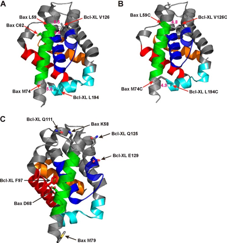
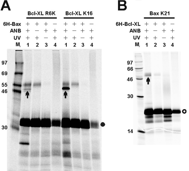
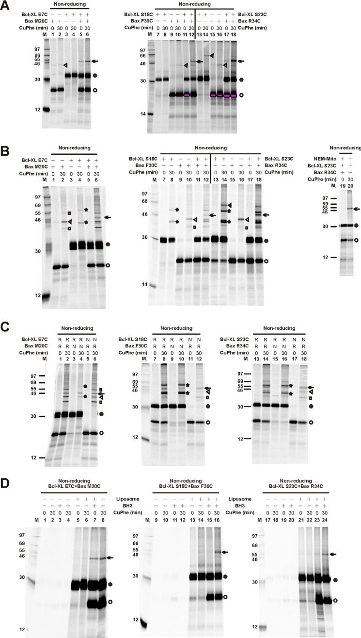
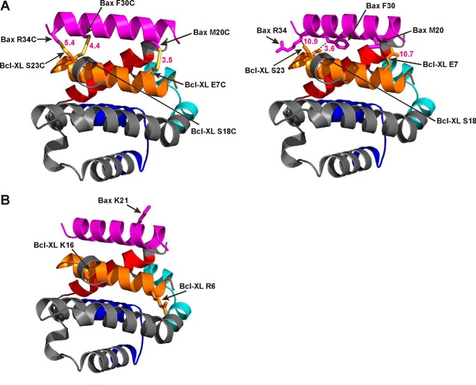
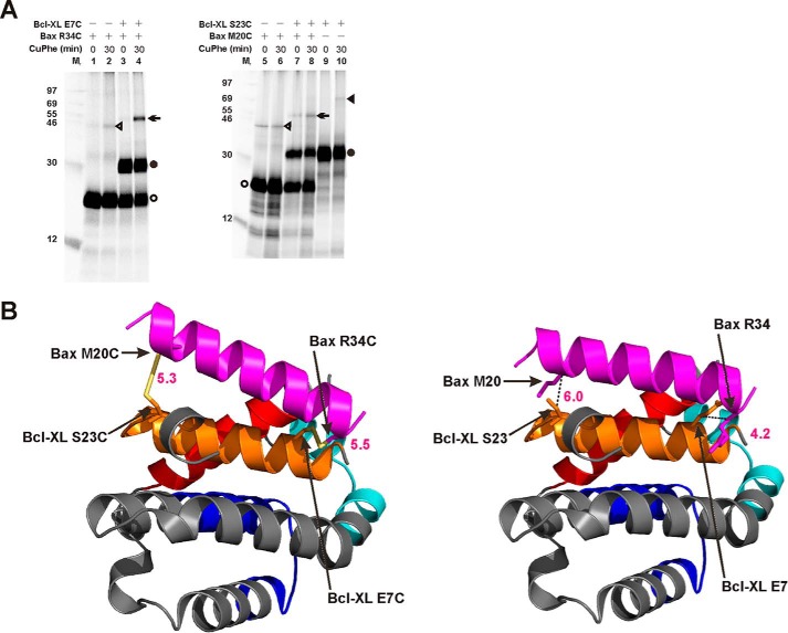
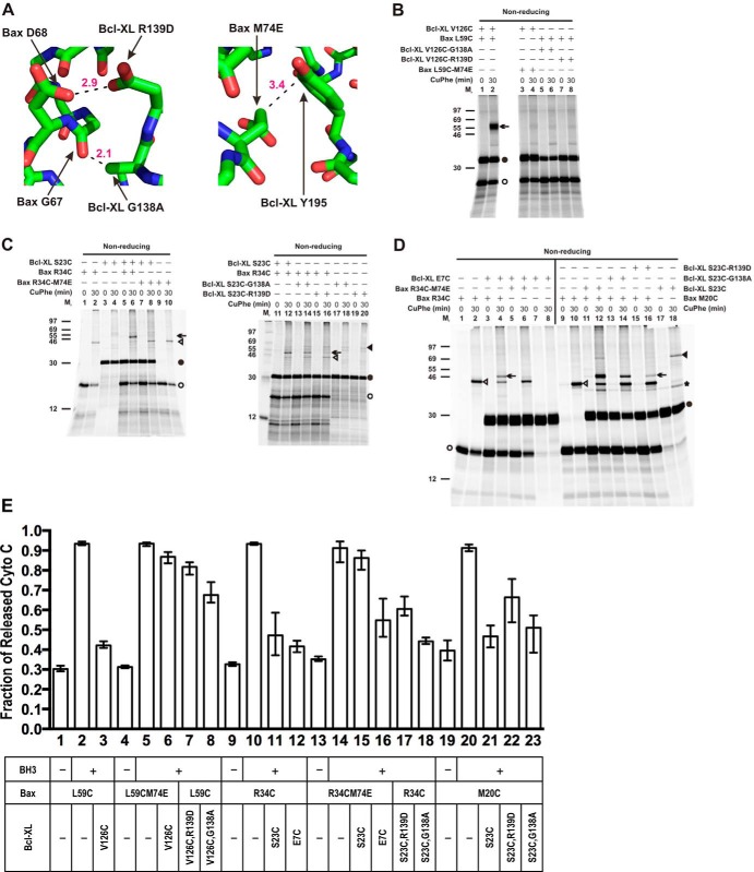
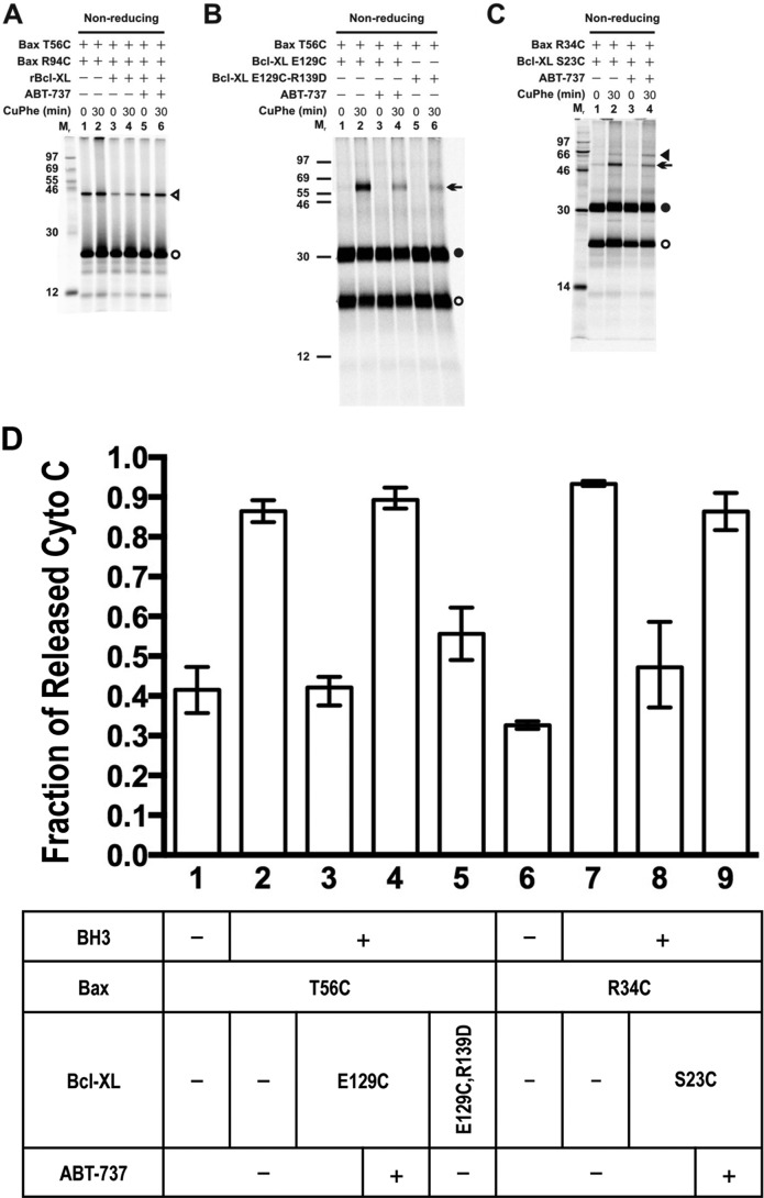
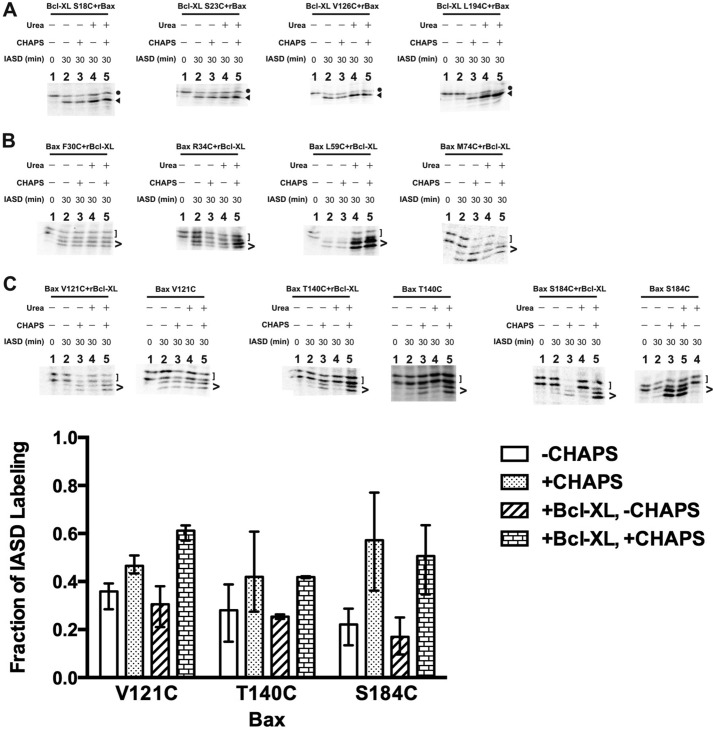
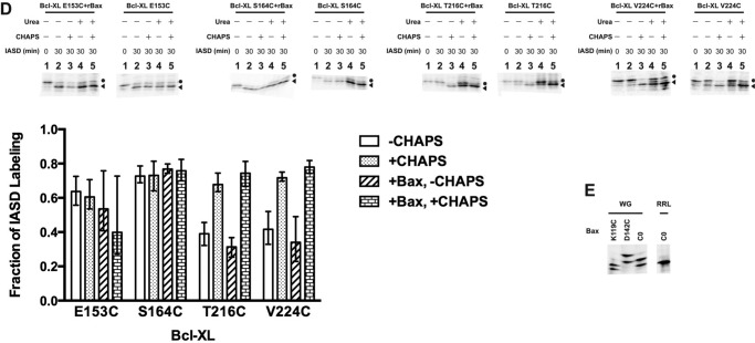
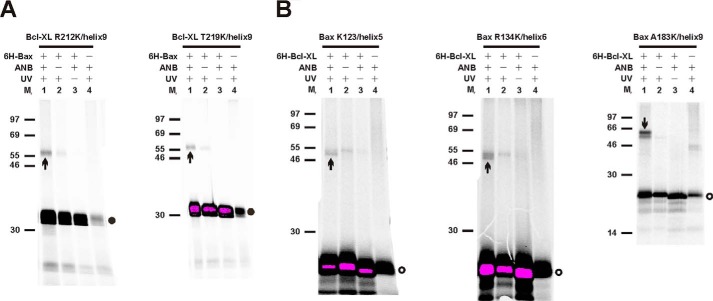
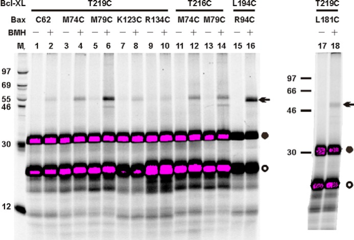
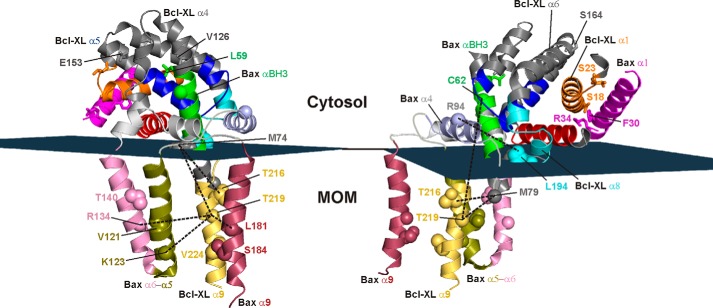
Bcl-XL binds to Bax, inhibiting Bax oligomerization required for mitochondrial outer membrane permeabilization (MOMP) during apoptosis. How Bcl-XL binds to Bax in the membrane is not known. Here, we investigated the structural organization of Bcl-XL·Bax complexes formed in the MOM, including the binding interface and membrane topology, using site-specific cross-linking, compartment-specific labeling, and computational modeling. We found that one heterodimer interface is formed by a specific interaction between the Bcl-2 homology 1-3 (BH1-3) groove of Bcl-XL and the BH3 helix of Bax, as defined previously by the crystal structure of a truncated Bcl-XL protein and a Bax BH3 peptide (Protein Data Bank entry 3PL7). We also discovered a novel interface in the heterodimer formed by equivalent interactions between the helix 1 regions of Bcl-XL and Bax when their helical axes are oriented either in parallel or antiparallel. The two interfaces are located on the cytosolic side of the MOM, whereas helix 9 of Bcl-XL is embedded in the membrane together with helices 5, 6, and 9 of Bax. Formation of the helix 1·helix 1 interface partially depends on the formation of the groove·BH3 interface because point mutations in the latter interface and the addition of ABT-737, a groove-binding BH3 mimetic, blocked the formation of both interfaces. The mutations and ABT-737 also prevented Bcl-XL from inhibiting Bax oligomerization and subsequent MOMP, suggesting that the structural organization in which interactions at both interfaces contribute to the overall stability and functionality of the complex represents antiapoptotic Bcl-XL·Bax complexes in the MOM.

Keywords: Bax; Bcl-2 Proteins; Bcl-XL; Membrane Proteins; Mitochondrial Apoptosis; Protein Cross-linking.


Figures

FIGURE 1.
FIGURE 1.
Sequences of Bax and Bcl-XL mutants. A and B, single-cysteine Bax and Bcl-XL mutants. Bax (A) and Bcl-XL (B) sequences are shown with BH motifs highlighted by dashed lines above, and helices are identified by arrows below. The native cysteines (underlined) were changed to alanine to create the cysteine-null (C0) mutants. Single-cysteine Bax and Bcl-XL mutants were created from the respective cysteine-null mutants by individually replacing the residues in boldface type with cysteine. Arrowheads, Met74 of Bax and Gly138 and Arg139 of Bcl-XL that were changed to glutamate, alanine, and aspartate in Bax M74E and Bcl-XL G138A and R139D mutants, respectively. C, single-lysine Bcl-XL mutants. Bcl-XL sequence is shown with BH motifs and helices indicated as in B. The native lysines (underlined) were changed to arginine to create the lysine-null (K0) mutant. Single-lysine Bcl-XL mutants were created from the lysine-null mutant by individually replacing the residues in boldface type with lysine.
FIGURE 2.
FIGURE 2.
Activities of Bax and Bcl-XL mutants in Bax−/−/Bak−/− mitochondria and mouse embryonic fibroblasts. A-C, MOMP activity in the Bax−/−/Bak−/− mitochondria. Wild-type (WT) or the indicated mutant Bax proteins were synthesized in vitro, activated by tBid protein, and targeted to the Bax−/−/Bak−/− mitochondria in the absence or presence of WT or the indicated mutant Bcl-XL proteins that were synthesized in vitro. Cytochrome c release from the mitochondria was measured using ELISA. Data shown are average fractions of cytochrome c release from two to three independent experiments with the ranges indicated by error bars. D, the apoptotic activity of WT, lysine-null, and single-lysine mutant Bax in the bax−/−/bak−/− MEFs. The MEF cells were infected with retrovirus that co-expressed Bax and GFP from the same message using the IRES sequence between the two coding regions and treated with etoposide of the indicated concentrations. The cells were examined for apoptosis by annexin V staining. Both panels show the fraction of adherent annexin V-positive cells in the GFP-positive (infected and expressing Bax; black bar) and GFP-negative (uninfected and not expressing Bax; white bar) populations. The type of Bax protein expressed by the virus that was used to infect the cells is indicated below the plot. GFP, control virus that expressed only GFP; WT, K0, K21, etc., virus that expressed GFP and wild-type, lysine-null, or single-lysine Bax, respectively. Data shown are the averages from eight independent replicates. For each replicate, a minimum of 356 cells/well (average = 1084, maximum = 2613) or 309 cells/well (average = 849, maximum = 1442) was analyzed for the 2 or 0.5 μm etoposide-treated cells, respectively. Error bars, S.D.
FIGURE 3.
FIGURE 3.
Disulfide cross-linking of Bax and Bcl-XL proteins with a single cysteine in BH3 helix and BH1–3 groove, respectively. A, the in vitro synthesized [35S]Met-labeled cysteine-null and single-cysteine Bax and Bcl-XL proteins were activated by the BH3 peptide and targeted to the MOM liposomes. The resulting proteoliposomes were isolated and oxidized by CuPhe for 30 min. NEM and EDTA were then added to stop the oxidation. For the 0 min controls, NEM and EDTA were added prior to the addition of CuPhe. The resulting samples were analyzed by non-reducing and reducing SDS-PAGE and phosphorimaging. B, the in vitro synthesized radioactive (R) and/or non-radioactive (N) Bax and Bcl-XL mutants were activated by the BH3 peptide and targeted to the MOM liposomes. The resulting proteoliposomes were processed and analyzed as described in A. C, the in vitro synthesized radioactive Bax and Bcl-XL mutant proteins were incubated in the absence or presence of the MOM liposomes and/or the BH3 peptide. The resulting proteoliposomes were processed and analyzed as described in A. D and E, in vitro synthesized radioactive Bax and Bcl-XL mutants were activated by tBid and targeted to the MOM liposomes (D) or activated by the BH3 peptide and targeted to the Bax−/−/Bak−/− mitochondria (E). The resulting membranes were isolated, oxidized, and analyzed as described in A. F, the in vitro synthesized radioactive (R) and/or non-radioactive (N) Bax and Bcl-XL mutant proteins were activated by the BH3 peptide and targeted to the Bax−/−/Bak−/− mitochondria. The resulting mitochondria were isolated, oxidized, and analyzed as described in A. In all panels, protein standards are indicated on the side of phosphorimages with their molecular masses (Mr). Arrows, disulfide-linked Bax·Bcl-XL heterodimers; open or filled circles, Bax or Bcl-XL monomers, respectively.
FIGURE 4.
FIGURE 4.
Photocross-linking of Bcl-XL or Bax mutant proteins with a single photoreactive ANB-lysine incorporated into the BH1–3 groove or BH3 helix, respectively. The in vitro synthesized [35S]Met-labeled Bcl-XL (A) or Bax (B) mutant proteins without lysine (K0) and with a single photoreactive ANB probe incorporated at the indicated lysine positions (F97K, M79K, etc.) were mixed with the purified 6H-Bax (A) or 6H-Bcl-XL (B) protein. The proteins were activated by the BH3 peptide and targeted to the MOM liposomes. The resulting proteoliposomes were isolated and photolyzed. The resulting photoadducts of the 6H-tagged and the 35S-labeled proteins were enriched and analyzed by reducing SDS-PAGE and phosphorimaging. Protein standards are indicated on the side of phosphorimages with Mr. Filled or open circle, [35S]Bcl-XL or [35S]Bax monomer, respectively. Arrow, photoadduct of [35S]Bcl-XL and 6H-Bax (A) or [35S]Bax and 6H-Bcl-XL (B).
FIGURE 5.
FIGURE 5.
A model for the BH1–3 groove·BH3 helix interface in the membrane-bound Bcl-XL·Bax heterodimer. A, the crystal structure of Bcl-XL protein·Bax BH3 peptide complex (PDB entry 3PL7) is shown with Val126/Leu59 and Leu194/Met74, the residue pairs that formed disulfide bonds when they were replaced with cysteine pairs (Fig. 3), presented in stick form, and their β-carbon atoms (C1β and C2β) linked by dashed lines with the distances in Å indicated. Cys62, a native cysteine in the BH3 region of Bax, is also presented in stick form with its Cβ linked to the Cβ of Bcl-XL Val126 and the distance in Å indicated. B, the two Bcl-XL/Bax residue pairs, Val126/Leu59 and Leu194/Met74, shown in A are replaced by two cysteine pairs, which are presented in stick form with their γ-sulfur atoms (S1γ and S2γ) linked by virtual bonds and the distances in Å indicated. The C1β-S1γ-S2γ-C2β dihedral angles about the V126C/L59C and L194C/M74C disulfide bonds are −52.2 and +22.0°, respectively. C, the crystal structure of the Bcl-XL protein·Bax BH3 peptide complex is shown with the residues that generated the heterodimer-specific photoadducts when replaced by ANB-lysine (Fig. 4) presented in stick form. In all panels, the BH1, BH2, BH3, and BH4 regions of Bcl-XL protein are colored blue, cyan, red, and orange, respectively, and the BH3 region of the Bax peptide is colored green.
FIGURE 6.
FIGURE 6.
Photocross-linking of Bax and Bcl-XL mutant proteins with single photoreactive ANB-lysine incorporated into their helix 1. The [35S]Met-labeled Bcl-XL (A) or Bax (B) mutant proteins with single photo-reactive ANB probe incorporated at the indicated lysine positions were synthesized in vitro and activated together with the purified 6H-Bax (A) or 6H-Bcl-XL (B) protein, respectively, by the BH3 peptide. The proteins were targeted to the MOM liposomes, processed, and analyzed as described in the legend to Fig. 4.
FIGURE 7.
FIGURE 7.
Disulfide cross-linking of Bax and Bcl-XL proteins with a single cysteine in their helix 1. A and B, the in vitro synthesized [35S]Met-labeled single-cysteine Bcl-XL and Bax proteins were activated by the BH3 peptide and targeted to the MOM liposomes (A) or the Bax−/−/Bak−/− mitochondria (B). The resulting membrane-bound proteins were processed and analyzed as described in the legend to Fig. 3. C, the in vitro synthesized radioactive (R) and/or non-radioactive (N) single-cysteine Bcl-XL and Bax proteins were activated by the BH3 peptide and targeted to the Bax−/−/Bak−/− mitochondria. The resulting mitochondria-bound proteins were processed and analyzed as in B. D, the in vitro synthesized radioactive single-cysteine Bcl-XL and Bax proteins were incubated in the absence or presence of the MOM liposomes and/or the BH3 peptide. The resulting proteoliposomes were processed and analyzed as in A. In all panels, the labels are as described in the legend to Fig. 1. In addition, filled or open triangles indicate the disulfide-linked Bcl-XL or Bax homodimers, and stars or squares indicate the disulfide-linked Bcl-XL or Bax·mitochondrial protein complexes, respectively. NEM-Mito in B indicates that the mitochondria used in the reaction were pretreated with NEM to block the sulfhydryl moieties in the mitochondrial proteins, preventing their cross-linking with the sulfhydryl moieties in the Bcl-XL and Bax proteins.
FIGURE 8.
FIGURE 8.
A model for the parallel helix 1·helix 1 dimer interface in the membrane-bound Bcl-XL·Bax heterodimer. A, left, to build the initial model, the helix 1 extracted from the Bax monomer structure (PDB entry 1F16) was manually positioned onto the helix 1 of the Bcl-XL protein in the Bcl-XL protein·Bax BH3 peptide complex structure (PDB entry 3PL7), such that the three cysteine pairs, E7C/M20C, S18C/F30C, and S23C/R34C, which resulted in disulfide-linked Bcl-XL·Bax heterodimers in Fig. 7, are in a geometry suitable for disulfide linkage. The resulting complex structure was input into FlexPepDock program, and one of the top 10 output models is shown with the cysteine pairs presented in stick form and their γ-sulfur atoms (S1γ and S2γ) linked by virtual bonds and the distances in Å indicated. The C1β-S1γ-S2γ-C2β dihedral angles about the disulfide bond for the three cysteine pairs are +99.2°, +114.5°, and −178.6°, respectively. Right, to generate the final model, the cysteines in the left panel were changed back to the corresponding wild-type residues. The resulting complex structure was the starting model in an automated peptide docking experiment with the FlexPepDock program. One of the top 10 output models from the docking experiment is shown with the respective wild-type residue pairs presented in stick form and their β-carbon atoms (C1β and C2β) linked by dashed lines with the distances in Å indicated. B, a model for the parallel helix 1·helix 1 interface in the Bcl-XL·Bax heterodimer was built based on the model presented in the right panel of A and the photocross-linking data shown in Fig. 6. The residues that generated the heterodimer-specific photoadducts when replaced by ANB-lysine are presented in stick form. In all models, the BH1–4 regions of Bcl-XL protein are colored as in Fig. 5, and the helix 1 of Bax is colored magenta. For simplicity, the BH3 helix of Bax was omitted from the models.
FIGURE 9.
FIGURE 9.
Disulfide cross-linking of Bax and Bcl-XL proteins with a single cysteine in their helix 1 that supports the antiparallel helix 1·helix 1 dimer model. A, the in vitro synthesized [35S]Met-labeled single-cysteine Bcl-XL and Bax proteins were activated by the BH3 peptide and targeted to the MOM liposomes. The resulting membrane-bound proteins were processed and analyzed as described in Fig. 7. B, the antiparallel helix 1·helix 1 dimer model was based on the disulfide cross-linking data from two single-cysteine Bcl-XL/Bax mutant pairs, E7C/R34C and S23C/M20C, shown in A. It was generated by the FlexPepDock program and presented similarly as described in the legend to Fig. 8A. Left, the two cysteine pairs are presented in stick form with their γ-sulfur atoms (S1γ and S2γ) linked by virtual bonds and the distances in Å indicated. The C1β-S1γ-S2γ-C2β dihedral angles about the disulfide bond for the two cysteine pairs are +146.7° and −74.0°, respectively. Right, the two respective wild-type residue pairs are presented in stick form with their β-carbon atoms (C1β and C2β) linked by dashed lines and the distances in Å indicated. The BH1–4 regions of Bcl-XL protein and the helix 1 of Bax are colored as in Fig. 8. For simplicity, the BH3 helix of Bax was omitted from the models.
FIGURE 10.
FIGURE 10.
Effects of interfacial mutations on Bcl-XL·Bax heterodimer formation and function. A, a part of the crystal structure of Bcl-XL protein·Bax BH3 peptide complex (PDB entry 3PL7) is shown in the left panel with Bcl-XL Gly138 changed to alanine (G138A) and Arg139 changed to aspartate (R139D) and in the right panel with Bax Met74 changed to glutamate (M74E). The mutated residues are shown in stick form. The residues in the respective binding partners that interact with these mutated residues are also illustrated in stick form with dashed lines linking the Cβ of Bcl-XL Ala138 to the backbone carbonyl O of Bax Gly67, Oδ1 of Bcl-XL Asp139 to Oδ1 of Bax Asp68, and the Cγ of Bax Glu74 to Cϵ2 of Bcl-XL Tyr195. The distances between these atoms are indicated in Å. B–D, the in vitro synthesized [35S]Met-labeled single-cysteine Bcl-XL and Bax proteins with or without the indicated interfacial mutations were activated by the BH3 peptide and targeted to the Bax−/−/Bak−/− mitochondria that were either untreated (B) or pretreated with NEM (C and D). The mitochondria-bound proteins were processed and analyzed as described in the legend to Fig. 3. E, the indicated Bax proteins were synthesized in vitro, and their BH3 peptide-dependent cytochrome c release activities in the Bax−/−/Bak−/− mitochondria were assayed in the absence or presence of the indicated in vitro synthesized Bcl-XL proteins as described in the legend to Fig. 2. Data shown are average fractions of cytochrome c release from 2–4 independent experiments with the ranges indicated by error bars.
FIGURE 11.
FIGURE 11.
Effects of ABT-737 on interactions and MOMP activity of Bcl-XL and Bax. A-C, the indicated in vitro synthesized [35S]Met-labeled single-cysteine Bax (A), Bax and Bcl-XL (B and C), or Bax and Bcl-XL R139D (B) proteins were activated by the BH3 peptide and targeted to the MOM liposomes in the absence or presence of ABT-737, purified non-radioactive recombinant Bcl-XL protein (rBcl-XL), or both. The resulting proteoliposomes were processed and analyzed as described in the legend to Fig. 3. D, cytochrome c release from the Bax−/−/Bak−/− mitochondria by the indicated single-cysteine Bax proteins synthesized in vitro was assayed in the absence or presence of the BH3 peptide, the indicated single-cysteine Bcl-XL protein synthesized in vitro, and/or ABT-737, as described in the legend to Fig. 2. Data shown are average fractions of cytochrome c release from 2–6 independent experiments with the ranges indicated by error bars.
FIGURE 12.
FIGURE 12.
IASD-labeling of single-cysteine Bax and Bcl-XL proteins in liposomal membranes. The [35S]Met-labeled mutants with a single cysteine positioned in helix 1 and BH1–3 groove of Bcl-XL (A), helix 1 and BH3 helix of Bax (B), or helices 5, 6, and 9 of Bax (C, top) or of Bcl-XL (D, top) were synthesized in the wheat germ-based in vitro system. The resulting radioactive Bcl-XL or Bax protein, either alone or together with the purified non-radioactive recombinant Bax (rBax) or Bcl-XL (rBcl-XL) protein, respectively, were activated by the BH3 peptide and targeted to the MOM liposomes. The resulting proteoliposomes were isolated and treated with IASD in the absence or presence of CHAPS, urea, or both. After 30 min, the labeling reactions were stopped by β-mercaptoethanol. For the 0 min controls, the samples were pretreated with β-mercaptoethanol before the addition of IASD. The resulting radioactive proteins were resolved using IEF and detected by phosphorimaging. Circles and triangles, unlabeled and IASD-labeled Bcl-XL proteins, respectively; square and angle brackets, unlabeled and IASD-labeled Bax, respectively. The phosphorimaging data for IASD labeling of radioactive Bcl-XL and Bax mutants in the top panels of C and D and the similar data from 1–4 independent replicates were quantified. In the corresponding bottom panels, the average fractions of IASD labeling for the mutants under the specified conditions are presented as bar graphs of the specified patterns with the ranges indicated by error bars. E, the indicated [35S]Met-labeled Bax mutants were synthesized in the wheat germ (WG)- or rabbit reticulocyte lysate (RRL)-based in vitro translation system and analyzed with IEF and phosphorimaging. The K119C or D142C mutant was constructed from the C0 mutant by changing Lys119 or Asp142 to cysteine, respectively.
FIGURE 12.
FIGURE 12.
IASD-labeling of single-cysteine Bax and Bcl-XL proteins in liposomal membranes. The [35S]Met-labeled mutants with a single cysteine positioned in helix 1 and BH1–3 groove of Bcl-XL (A), helix 1 and BH3 helix of Bax (B), or helices 5, 6, and 9 of Bax (C, top) or of Bcl-XL (D, top) were synthesized in the wheat germ-based in vitro system. The resulting radioactive Bcl-XL or Bax protein, either alone or together with the purified non-radioactive recombinant Bax (rBax) or Bcl-XL (rBcl-XL) protein, respectively, were activated by the BH3 peptide and targeted to the MOM liposomes. The resulting proteoliposomes were isolated and treated with IASD in the absence or presence of CHAPS, urea, or both. After 30 min, the labeling reactions were stopped by β-mercaptoethanol. For the 0 min controls, the samples were pretreated with β-mercaptoethanol before the addition of IASD. The resulting radioactive proteins were resolved using IEF and detected by phosphorimaging. Circles and triangles, unlabeled and IASD-labeled Bcl-XL proteins, respectively; square and angle brackets, unlabeled and IASD-labeled Bax, respectively. The phosphorimaging data for IASD labeling of radioactive Bcl-XL and Bax mutants in the top panels of C and D and the similar data from 1–4 independent replicates were quantified. In the corresponding bottom panels, the average fractions of IASD labeling for the mutants under the specified conditions are presented as bar graphs of the specified patterns with the ranges indicated by error bars. E, the indicated [35S]Met-labeled Bax mutants were synthesized in the wheat germ (WG)- or rabbit reticulocyte lysate (RRL)-based in vitro translation system and analyzed with IEF and phosphorimaging. The K119C or D142C mutant was constructed from the C0 mutant by changing Lys119 or Asp142 to cysteine, respectively.
FIGURE 13.
FIGURE 13.
Photocross-linking of Bcl-XL and Bax proteins with a single photoreactive ANB-lysine incorporated into the membrane-embedded helices. The in vitro synthesized [35S]Met-labeled Bcl-XL (A) or Bax (B) protein with the photoreactive probe attached to the indicated single lysine in helix 5, 6, or 9 was activated together with purified 6H-Bax (A) or 6H-Bcl-XL (B) protein, respectively, by the BH3 peptide and targeted to the MOM liposomes. The resulting proteoliposomes were processed and analyzed as described in the legend to Fig. 4.
FIGURE 14.
FIGURE 14.
Chemical cross-linking of single-cysteine Bcl-XL and Bax proteins. The indicated in vitro synthesized [35S]Met-labeled single-cysteine Bcl-XL and Bax proteins were activated by the BH3 peptide and targeted to the MOM liposomes. The resulting proteoliposomes were isolated and subjected to BMH cross-linking. The resulting radioactive proteins and their adducts were analyzed by reducing SDS-PAGE and phosphorimaging. Arrows, BMH-cross-linked heterodimers. Other labels are the same as those in Fig. 3.
FIGURE 15.
FIGURE 15.
A model for the overall structural organization of the Bcl-XL·Bax heterodimer in membranes. The cytosolic part of the heterodimer model was assembled by merging the BH1–3 groove·BH3 helix interface model (Fig. 5A) with the parallel helix 1·helix 1 interface model (Fig. 8A, right). The MOM-embedded part was assembled with the α-helix 9 (α9) of Bcl-XL and the α-helices 5, 6, and 9 (α5, α6, and α9) of Bax. The distances between these membrane-embedded helices were set according to the BMH cross-linking data (Fig. 14). The distances between the helix α8 or α9 of Bcl-XL and the BH3 helix (αBH3) or helix α4 of Bax, respectively, were further adjusted based on the BMH cross-linking data. Dashed lines link the residues that were cross-linked by BMH when they were mutated to cysteines. The residues shown as spheres are embedded in the MOM, because their cysteine substitutions could not be substantially labeled by IASD unless CHAPS was added (Fig. 12). The residues shown as sticks are exposed to the cytosol, because their cysteine substitutions could be labeled by IASD in the absence of CHAPS and urea.

Similar articles

Cited by

References

    1. Kvansakul M., Yang H., Fairlie W. D., Czabotar P. E., Fischer S. F., Perugini M. A., Huang D. C., Colman P. M. (2008) Vaccinia virus anti-apoptotic F1L is a novel Bcl-2-like domain-swapped dimer that binds a highly selective subset of BH3-containing death ligands. Cell Death Differ. 15, 1564–1571 - PubMed
    1. Youle R. J., Strasser A. (2008) The BCL-2 protein family: opposing activities that mediate cell death. Nat. Rev. Mol. Cell Biol. 9, 47–59 - PubMed
    1. Leber B., Lin J., Andrews D. W. (2007) Embedded together: the life and death consequences of interaction of the Bcl-2 family with membranes. Apoptosis 12, 897–911 - PMC - PubMed
    1. Chipuk J. E., Moldoveanu T., Llambi F., Parsons M. J., Green D. R. (2010) The BCL-2 family reunion. Mol. Cell 37, 299–310 - PMC - PubMed
    1. Billen L. P., Kokoski C. L., Lovell J. F., Leber B., Andrews D. W. (2008) Bcl-XL inhibits membrane permeabilization by competing with Bax. PLoS Biol. 6, e147. - PMC - PubMed

Publication types

Substances

LinkOut - more resources