. 2014 Apr 25;289(17):11571-11583.
doi: 10.1074/jbc.M113.546168. Epub 2014 Mar 10.

Crystal structures of the human RNA demethylase Alkbh5 reveal basis for substrate recognition

Affiliations

Crystal structures of the human RNA demethylase Alkbh5 reveal basis for substrate recognition

Chong Feng et al. J Biol Chem. .

Abstract

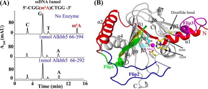
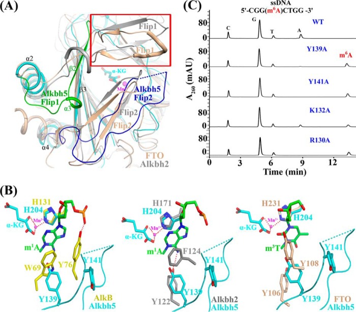
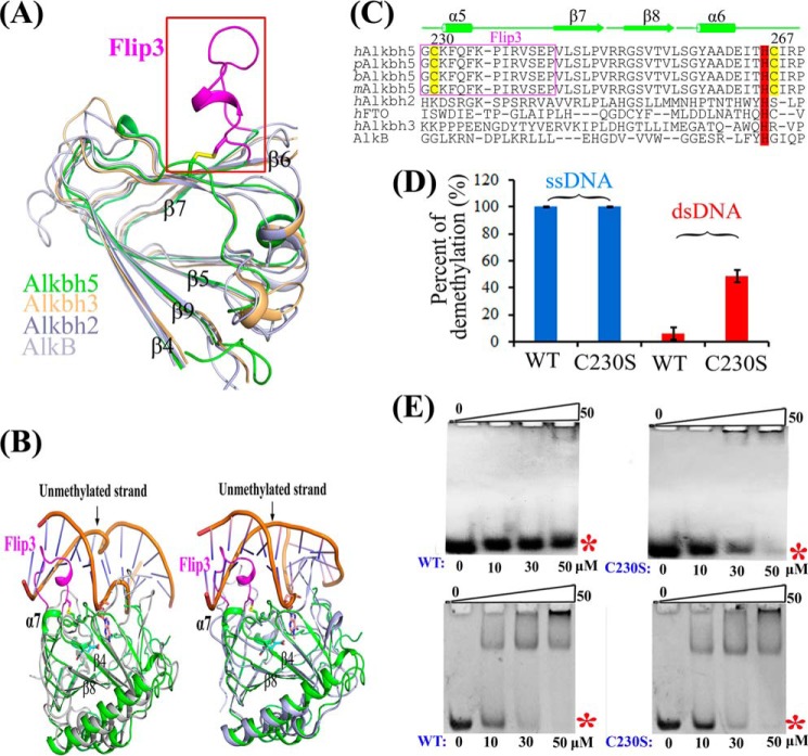
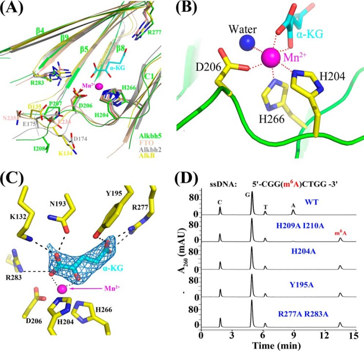
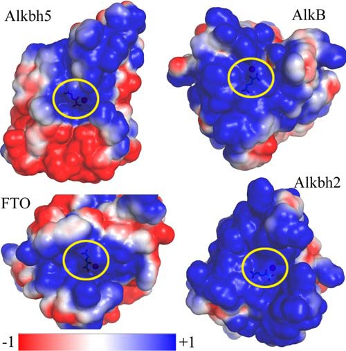
N(6)-Methylation of adenosine is the most ubiquitous and abundant modification of nucleoside in eukaryotic mRNA and long non-coding RNA. This modification plays an essential role in the regulation of mRNA translation and RNA metabolism. Recently, human AlkB homolog 5 (Alkbh5) and fat mass- and obesity-associated protein (FTO) were shown to erase this methyl modification on mRNA. Here, we report five high resolution crystal structures of the catalytic core of Alkbh5 in complex with different ligands. Compared with other AlkB proteins, Alkbh5 displays several unique structural features on top of the conserved double-stranded β-helix fold typical of this protein family. Among the unique features, a distinct "lid" region of Alkbh5 plays a vital role in substrate recognition and catalysis. An unexpected disulfide bond between Cys-230 and Cys-267 is crucial for the selective binding of Alkbh5 to single-stranded RNA/DNA by bringing a "flipping" motif toward the central β-helix fold. We generated a substrate binding model of Alkbh5 based on a demethylation activity assay of several structure-guided site-directed mutants. Crystallographic and biochemical studies using various analogs of α-ketoglutarate revealed that the active site cavity of Alkbh5 is much smaller than that of FTO and preferentially binds small molecule inhibitors. Taken together, our findings provide a structural basis for understanding the substrate recognition specificity of Alkbh5 and offer a foundation for selective drug design against AlkB members.

Keywords: AlkB; Alkbh5; Crystal Structure; Drug Design; Enzyme Inhibitors; RNA Modification; Substrate Recognition; m6A Demethylase; mRNA.


Figures

FIGURE 1.
FIGURE 1.
Overall structure of human Alkbh5·α-KG·Mn2+. A, Alkbh5 in vitro enzymatic activity assays. As shown by the HPLC analysis of digested substrates, Alkbh5 66–292 used for crystallization shows the same repair activity as the longer fragment Alkbh5 66–394. B, the overall structures of Alkbh5 66–292 in the presence of α-KG and Mn2+. The Mn2+ ion is shown as a magenta sphere. The disulfide bond between Cys-230 and Cys-267 is highlighted in yellow and indicated by the black arrow. Key residues as well as α-KG are shown as sticks, and secondary structures are labeled. Flip1, Flip2, and Flip3 are shown in green, blue, and magenta, respectively. mAU, milli-absorbance units.
FIGURE 2.
FIGURE 2.
The distinct lid region of Alkbh5 is vital for its substrate recognition and catalysis. A, structural comparison of Alkbh5 with FTO (tinted; Protein Data Bank code 3LFM) and Alkbh2 (gray; Protein Data Bank code 3BTZ) around the lid region. The most distinct regions, Flip1 (residues 117–129) and Flip2 (residues 136–165), of Alkbh5 are shown in green and blue, respectively. The discrete region is shown in blue dashed lines. The uncovered and larger space over the active site of Alkbh5 is labeled in the red frame. B, superposition of the partial key residues involved in nucleoside recognition. Alkbh5 (cyan), AlkB (Protein Data Bank code 3BIE; yellow; left panel), Alkbh2 (Protein Data Bank code 3BTZ; gray; middle panel), and FTO (Protein Data Bank code 3LFM; tinted; right panel) are shown. The nucleoside is colored green. Hydrogen bonds are indicated as red dashed lines. α-KG and Mn2+ from Alkbh5 are labeled. C, mutations of the key residues in the lid region (shown in supplemental Movie S1) greatly impair Alkbh5 demethylation activity. mAU, milli-absorbance units.
FIGURE 3.
FIGURE 3.
The unique disulfide bond of Alkbh5 decides its binding preference for single-stranded nucleic acids. A, structural comparison of Alkbh5 with Alkbh3 (tinted; Protein Data Bank code 2IUW), Alkbh2 (blue; Protein Data Bank code 3BTZ), and AlkB (gray; Protein Data Bank code 3BIE) around the jelly roll motif. The unique Flip3 (magenta; residues 229–242) of Alkbh5 is highlighted within the red frame. B, structural alignment of Alkbh5 with AlkB·dsDNA complex (left) and Alkbh2·dsDNA complex (right). The unmethylated strand of dsDNA would sterically clash with “Flip3” of Alkbh5. 1-meA (purple) from the methylated strand of dsDNA and α-KG (cyan) from Alkbh5 are shown in sticks. C, structure-based sequence alignment of Alkbh5 of different species and its family members within the disulfide bond-forming region. The conserved Fe2+-binding residue His-266 is colored in red. h, Homo sapiens; p, Pan troglodytes; b, Bos taurus; m, Mus musculus. D, comparison of the demethylation activity of WT and C230S mutant Alkbh5 for the single-stranded and double-stranded m6A-containing oligonucleotides. All experiments were repeated three times. Error bars indicate ±S.D. E, EMSA binding assays were performed for dsDNA (upper panel) and partial duplex DNA with a 5′ eight-nucleotide ss-DNA (lower panel) with increasing amounts (indicated at the bottom) of Alkbh5 WT (left) and C230S mutant (right). The sequence of the partial duplex DNA with a 5′ eight-nucleotide ssDNA was as follows: 5′-CGGACTGGCGGCAGCACTGC-3′ and 5′-GCAGTGCTGCCG-3′. Total DNA substrate used was 10 μm. The position of free DNA is indicated by a red asterisk.
FIGURE 4.
FIGURE 4.
The detailed interaction of the active center of Alkbh5. A, the superposition of Alkbh5 (green) with AlkB (yellow), Alkbh2 (gray), and FTO (tinted) at the active site. All the critical residues are shown as sticks. The two residues following the conserved HX(D/E) motif from the four proteins, α-KG, and Mn2+ from Alkbh5 are labeled. B, the interaction network around Mn2+. Mn2+, water, α-KG, and the side chains of Alkbh5 are colored in magenta, blue, cyan, and yellow, respectively. Interaction between Mn2+ and its coordinating atoms are shown in red dashed lines. C, the interaction network around α-KG and the involved residues are shown as sticks. The FoFc differential electron density map (contour level, σ = 3.0) is indicated as a marine blue mesh. D, demethylation activity to m6A-containing ssDNA of the wide-type Alkbh5 and its mutants involved in the Mn2+ binding, α-KG binding, and the residues around the entrance loop of the jelly roll fold. mAU, milli-absorbance units.
FIGURE 5.
FIGURE 5.
The substrate recognition and potential binding model of Alkbh5. α-KG and Mn2+ are shown. A, the electrostatic surface potential of Alkbh5. The positively charged surface is colored blue, and the negatively charged surface is colored red. The two positively charged grooves around Flip3 are highlighted and labeled. The key residues are shown as sticks and labeled. B, the critical residues located at the positively charged surface of Alkbh5 are presented. The view orientation is the same as A. The residues in the missing part of Flip2 are shown in blue dashed lines. C, repair activity of Alkbh5 surface residue variants on the 8-mer m6A-containing ssDNA. Error bars indicate ±S.D. for triplicate experiments. D, Alkbh5 shows no repair activity for 6-methyldeoxyadenosine as detected by HPLC. E, the proposed model of Alkbh5 binding to ssRNA (yellow). The m6A nucleobase inserted into the catalytic pocket is indicated, and Flip2 and Flip3 of Alkbh5 are also labeled. The key residues are shown as sticks; residues Lys-231, Lys-235, His-209, and Ile-210 are also labeled. mAU, milli-absorbance units.
FIGURE 6.
FIGURE 6.
Comparison of electrostatic surfaces among Alkbh5, AlkB, FTO, and Alkbh2. All the structures are shown in the same scale. Positively charged surface is colored in blue, negatively charged surface is colored in red, and neutral surface is colored in white. Alkbh5 is less positively charged compared with AlkB (Protein Data Bank code 3BIE), FTO (Protein Data Bank code 3LFM), and Alkbh2 (Protein Data Bank code 3BTZ). α-KG and Mn2+ are shown. The active center is shown in a yellow ellipse.
FIGURE 7.
FIGURE 7.
The binding of different ligands to Alkbh5. Citrate/acetate, α-KG, NOG, and PDCA are shown in green, cyan, magenta, and pale yellow, respectively. A, the interaction network around the citrate and acetate molecules in the naturally grown crystal structure of Alkbh5. Also the FoFc differential electron density map of the bound citrate and acetate at 3 σ is presented. Hydrogen bonds are indicated as black dashes. B, the superimposition of the different ligands at the active sites of Alkbh5·cofactor complexes. The greatest difference lies in residue Asp-206 of the Alkbh5·citrate complex; it rotates by 86.8° in the other three structures. Only the key residues from the Alkbh5·citrate complex and Alkbh5·α-KG complex are labeled. C, IC50 curves for inhibitors (inh), including succinate, PDCA, NOG, and citrate. Succinate is colored in blue. Error bars indicate ±S.D. D, superimposition of Alkbh5 (cyan) and FTO (gray; Protein Data Bank code 4IE7) around the binding pocket. Considerable steric hindrance (shown in cyan lines) between residues Ile-281 and Tyr-195 of Alkbh5 with the citrate would be introduced if citrate in Alkbh5 is bound like that in FTO·citrate. E, quantification of the binding affinity of α-KG (left) and succinate (right) to Alkbh5 by ITC. The integrated heat is plotted against the molar ratio of ligand added to Alkbh5 in the cell. The dissociation constant (Kd) is indicated. F, the interaction network around NOG (left) and PDCA (right). The residues involved are shown as sticks. The FoFc differential electron density maps (contour level, σ = 3.0) of NOG and PDCA are both indicated as a lime green mesh.

Similar articles

Cited by

References

    1. Cantara W. A., Crain P. F., Rozenski J., McCloskey J. A., Harris K. A., Zhang X., Vendeix F. A., Fabris D., Agris P. F. (2011) The RNA Modification Database, RNAMDB: 2011 update. Nucleic Acids Res. 39, D195–D201 - PMC - PubMed
    1. Wei C., Gershowitz A., Moss B. (1975) N6,O2′-dimethyladenosine a novel methylated ribonucleoside next to the 5′ terminal of animal cell and virus mRNAs. Nature 257, 251–253 - PubMed
    1. Canaani D., Kahana C., Lavi S., Groner Y. (1979) Identification and mapping of N6-methyladenosine containing sequences in simian virus 40 RNA. Nucleic Acids Res. 6, 2879–2899 - PMC - PubMed
    1. Harper J. E., Miceli S. M., Roberts R. J., Manley J. L. (1990) Sequence specificity of the human mRNA N6-adenosine methylase in vitro. Nucleic Acids Res. 18, 5735–5741 - PMC - PubMed
    1. Meyer K. D., Saletore Y., Zumbo P., Elemento O., Mason C. E., Jaffrey S. R. (2012) Comprehensive analysis of mRNA methylation reveals enrichment in 3′ UTRs and near stop codons. Cell 149, 1635–1646 - PMC - PubMed

Publication types