. 2014 Apr;9(4):823-32.
doi: 10.1002/cmdc.201300480. Epub 2014 Feb 24.

Crystal structure of human soluble adenylate cyclase reveals a distinct, highly flexible allosteric bicarbonate binding pocket

Affiliations

Crystal structure of human soluble adenylate cyclase reveals a distinct, highly flexible allosteric bicarbonate binding pocket

Susanne M Saalau-Bethell et al. ChemMedChem. 2014 Apr.

Abstract

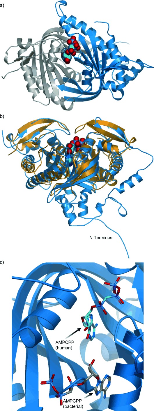
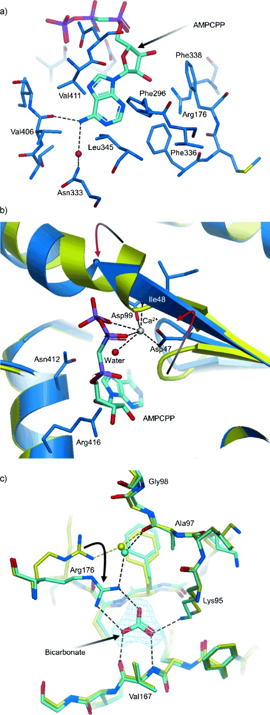
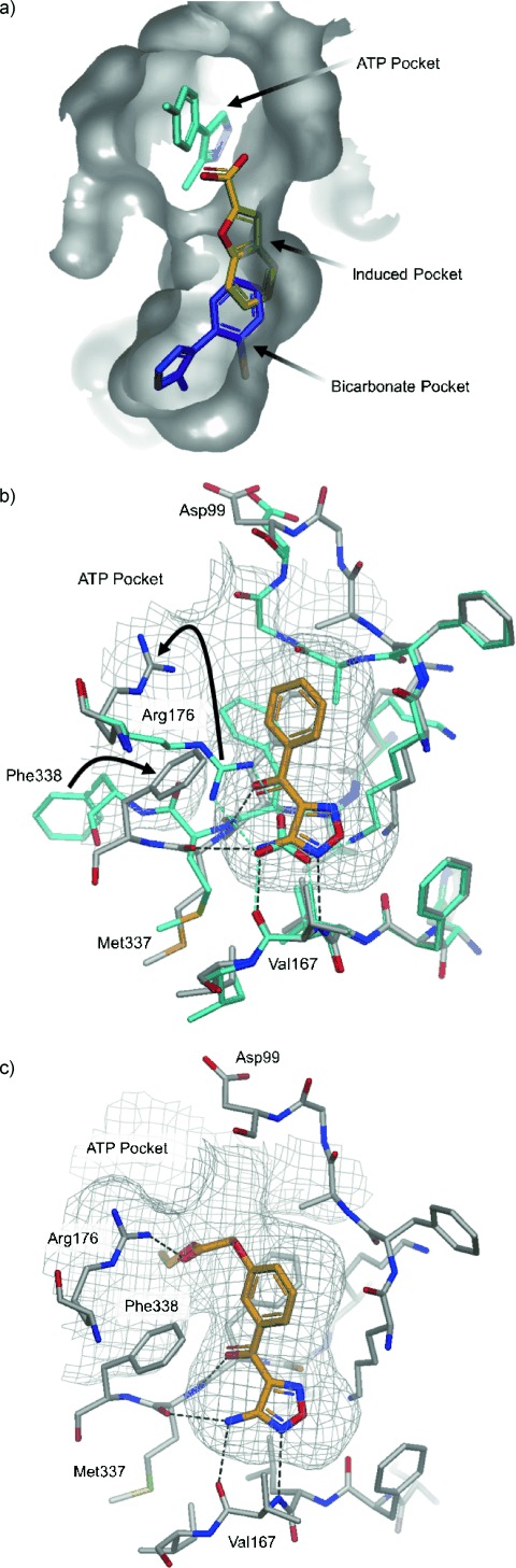
Soluble adenylate cyclases catalyse the synthesis of the second messenger cAMP through the cyclisation of ATP and are the only known enzymes to be directly activated by bicarbonate. Here, we report the first crystal structure of the human enzyme that reveals a pseudosymmetrical arrangement of two catalytic domains to produce a single competent active site and a novel discrete bicarbonate binding pocket. Crystal structures of the apo protein, the protein in complex with α,β-methylene adenosine 5'-triphosphate (AMPCPP) and calcium, with the allosteric activator bicarbonate, and also with a number of inhibitors identified using fragment screening, all show a flexible active site that undergoes significant conformational changes on binding of ligands. The resulting nanomolar-potent inhibitors that were developed bind at both the substrate binding pocket and the allosteric site, and can be used as chemical probes to further elucidate the function of this protein.

Keywords: allosterism; drug discovery; enzyme regulation; fragment screening; structural biology.


Figures

Figure 1
Figure 1
Crystal structure of the human adenylate cyclase. a) Ribbon diagram of the human soluble adenylate cyclase enzyme in complex with the substrate analogue AMPCPP (solid spheres). Domains C1 and C2 of the single chain are coloured blue and grey, respectively, and show the twofold pseudosymmetry of the protein. b) Overlay of the ribbon diagrams of the hsolAC–AMPCPP complex (blue) and the cyanobacterial solAC homodimer from Spirulina platensis (gold) showing the N-terminus and interdomain extensions of the human enzyme. The bound AMPCPP in hsolAC illustrates the location of the single active site. c) View of the hsolAC active site with bound AMPCPP (blue), with a molecule of AMPCPP (grey) from the S. platensis complex (PDB: 1WC0[17]) superimposed on the putative second site. The ribbon diagram of the human structure shows how the extension of β-strands 2′ and 3′ and the loop linking them (residues Met 337–Gly 341) occludes the site, obstructing the binding of a second nucleotide.
Figure 2
Figure 2
Close up views of the nucleotide and bicarbonate binding sites. a) hsolAC active site in complex with AMPCPP. The adenine pocket is shaped by hydrophobic residues Val 411, Phe 336, Leu 345, Phe 296 (Ala 97 is towards the back and not shown). The hydrogen bonds of the N6 amine to Val 406 backbone carbonyl and a water molecule (red sphere) are represented by dashed lines. Phe 338 and Arg 176 flank the ribose ring. b) Comparison between the apo (yellow) and AMPCPP–Ca2+ complex (blue) structures of hsolAC. The highly conserved Asn 412 and Arg 416 of the NXXXR cyclase catalytic motif hydrogen bond to the ribose and the phosphate oxygens. The Ca2+ ion (silver sphere) exhibits an octahedral coordination with β,γ oxygen atoms of the AMPCPP phosphates, a carboxyl oxygen each from the invariant Asp 47 and Asp 99 residues, the backbone carbonyl of Ile 48 and a water molecule (red sphere). The arrows highlight the movement of the β-hairpin loop and the unwinding of helix α3. c) Comparison of the apo (yellow) and bicarbonate-bound (cyan) hsolAC structures. Density for bicarbonate is shown (blue mesh). The bicarbonate anion forms hydrogen bonds (black dashed lines) with the backbone carbonyl and NH groups of Val 167, the guanidino group of Arg 176 and amine side chain of Lys 95. The side chain of Arg 176 undergoes an approximate 30° swing to hydrogen bond with bicarbonate, altering the water-mediated interaction with Ala 97.
Figure 3
Figure 3
Fragment binding and structure-guided compound optimisation. a) Overlay of three different crystal structures showing fragments binding in multiple regions of the hsolAC catalytic and allosteric pockets. The phenyl pyrazole (cyan) binds in the ATP pocket, an aromatic carboxylic acid (2; yellow) induces a new pocket and another pyrazole (3; magenta) binds in the allosteric bicarbonate pocket. b) Overlay of the bicarbonate (cyan) and amino-furazane fragment (4; orange) protein structures. Both molecules interact with the Val 167 backbone. Compound 4 triggers movement of the side chains of Arg 176 (close to the position the residue adopts in the apo structure) and Phe 338, altering the shape and electrostatic environment of the pocket. The ketone group of compound 4 forms a hydrogen bond with the backbone NH of Met 337, whilst the carbonyl of Met 337 forms an additional hydrogen bond with compound 4. The phenyl ring of the fragment sits in a hydrophobic pocket between Phe 45 and Phe 336 (not labelled, shown faded on the back). Position 3 on the phenyl ring of compound 4 presents a good vector to access the ATP pocket. c) Complex of hsolAC with compound 7 (orange). The ethyl ester added to the 3-methoxy group of compound 6 exploits the movement of Arg 176 observed with compound 4 (panel b) forming an additional hydrogen bond with its side chain and shows opportunity to further extend the inhibitors into the ATP pocket. d) Overlay of the protein complex of hsolAC with compounds 4 (cyan) and 8 (orange) showing the growth of the molecule from the original fragment. Interactions with both Val 167 and Met 337 backbones are preserved along the series. The molecule extends towards the ATP pocket forming two further hydrogen bonds: one with Arg 176 (also seen in the complex with compound 7) and an additional one, accessible through the movement of the Asp 99 side chain.
Scheme 1
Scheme 1
Reagents and conditions: a) DMF⋅DMA, toluene, reflux, 18 h; b) N2H4⋅H2O, EtOH, N2, reflux, 72 h.
Scheme 2
Scheme 2
Reagents and conditions: a) HNO3, AcOH, NaNO2, 60 °C, 2 h; b) NH3, Et2O, MeOH, 60 °C, 2 h.
Scheme 3
Scheme 3
Reagents and conditions: a) HNO3, AcOH, NaNO2, 60 °C, 2 h; b) NH3, Et2O, MeOH, 60 °C, 2 h; c) BCl3, CH2Cl2, RT, 30 min; d) NaH, DMF, 90 °C, 2 h.
Scheme 4
Scheme 4
Reagents and conditions: a) NaH, DMF, 75 °C, o/n.

References

    1. Casey JR. Biochem. Cell Biol. 2006;84:930–939. - PubMed
    1. Wutke MS, Buck J, Levin LR. JOP. 2001;2:154–158. - PubMed
    1. Buck J, Sinclair ML, Schapal L, Cann MJ, Levin LR. Proc. Natl. Acad. Sci. USA. 1999;96:79–84. - PMC - PubMed
    1. Chen Y, Cann MJ, Litvin TN, Iourgenko V, Sinclair ML, Levin LR, Buck J. Science. 2000;289:625–628. - PubMed
    1. Kamenetsky M, Middelhaufe S, Bank EM, Levin LR, Buck J, Steegborn C. J. Mol. Biol. 2006;362:623–639. - PMC - PubMed

MeSH terms

LinkOut - more resources