Review
. 2014 Feb 25:8:55.
doi: 10.3389/fncel.2014.00055. eCollection 2014.

Integration and regulation of glomerular inhibition in the cerebellar granular layer circuit

Affiliations
Review

Integration and regulation of glomerular inhibition in the cerebellar granular layer circuit

Lisa Mapelli et al. Front Cell Neurosci. .

Erratum in

Abstract

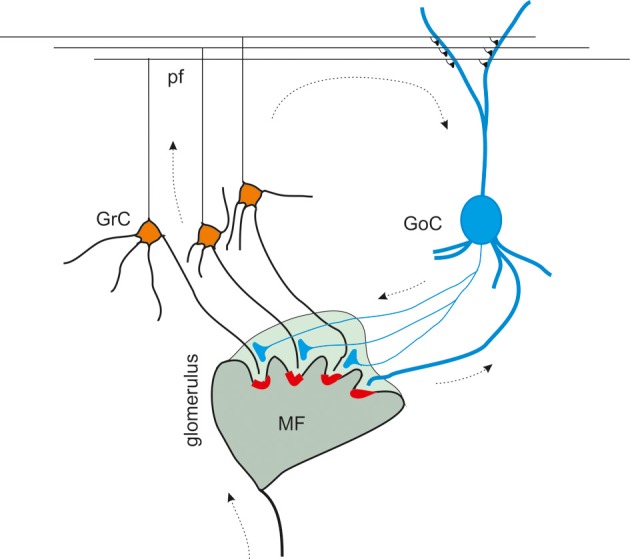
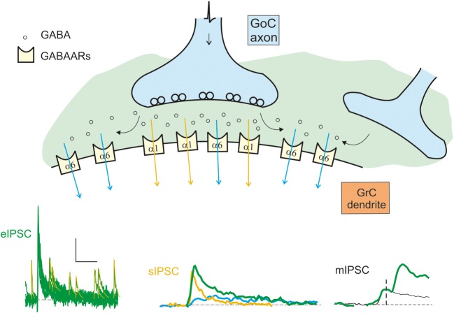
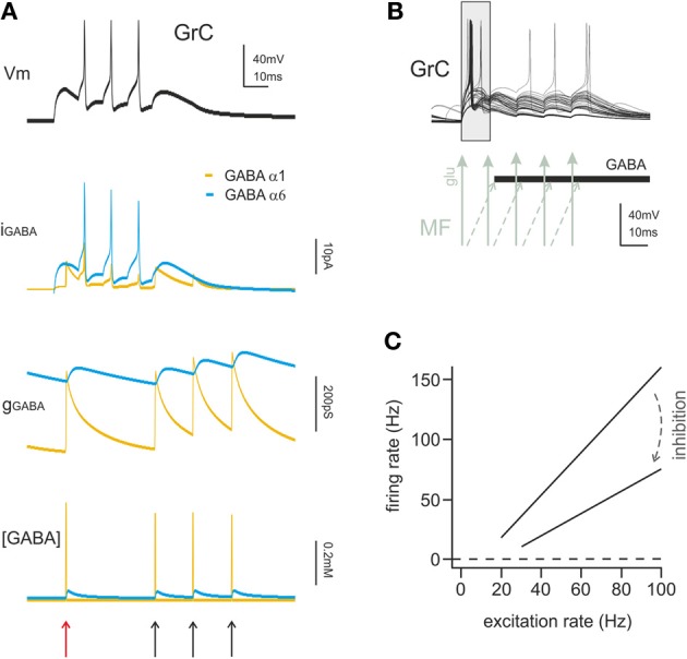
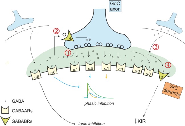
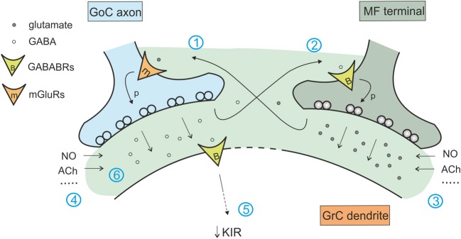
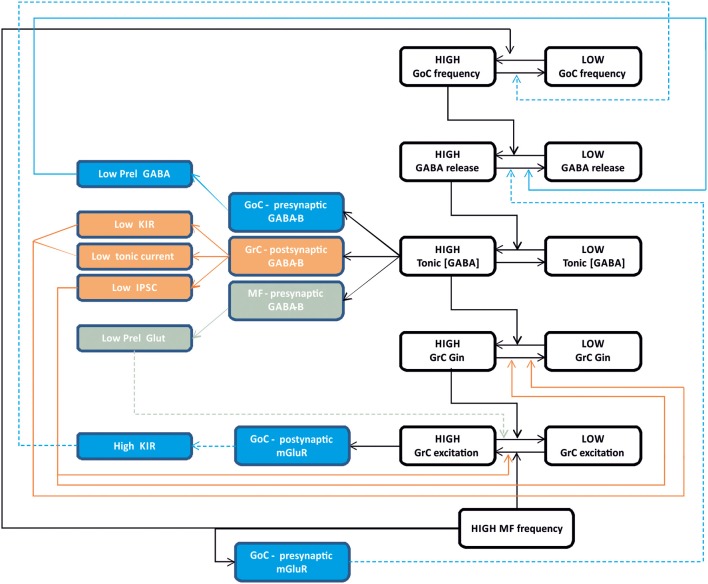
Inhibitory synapses can be organized in different ways and be regulated by a multitude of mechanisms. One of the best known examples is provided by the inhibitory synapses formed by Golgi cells onto granule cells in the cerebellar glomeruli. These synapses are GABAergic and inhibit granule cells through two main mechanisms, phasic and tonic. The former is based on vesicular neurotransmitter release, the latter on the establishment of tonic γ-aminobutyric acid (GABA) levels determined by spillover and regulation of GABA uptake. The mechanisms of post-synaptic integration have been clarified to a considerable extent and have been shown to differentially involve α1 and α6 subunit-containing GABA-A receptors. Here, after reviewing the basic mechanisms of GABAergic transmission in the cerebellar glomeruli, we examine how inhibition controls signal transfer at the mossy fiber-granule cell relay. First of all, we consider how vesicular release impacts on signal timing and how tonic GABA levels control neurotransmission gain. Then, we analyze the integration of these inhibitory mechanisms within the granular layer network. Interestingly, it turns out that glomerular inhibition is just one element in a large integrated signaling system controlled at various levels by metabotropic receptors. GABA-B receptor activation by ambient GABA regulates glutamate release from mossy fibers through a pre-synaptic cross-talk mechanisms, GABA release through pre-synaptic auto-receptors, and granule cell input resistance through post-synaptic receptor activation and inhibition of a K inward-rectifier current. Metabotropic glutamate receptors (mGluRs) control GABA release from Golgi cell terminals and Golgi cell input resistance and autorhythmic firing. This complex set of mechanisms implements both homeostatic and winner-take-all processes, providing the basis for fine-tuning inhibitory neurotransmission and for optimizing signal transfer through the cerebellar cortex.

Keywords: GABA receptors; Golgi cells; cerebellum; granule cells; synaptic inhibition.


Figures

Figure 1
Figure 1
The inhibitory circuit of cerebellar granular layer. In the granular layer of the cerebellar cortex, granule cells receive excitatory inputs from the mossy fibers and are inhibited by Golgi cells. The synaptic contacts among granule cell (GrC) dendrites, mossy fiber (MF) terminals, and Golgi cell (GoC) axon and dendrites are enwrapped into a glial sheet, originating a peculiar anatomical structure known as cerebellar glomerulus. Each glomerulus is characterized by one mossy fiber rosette and several granule cell and Golgi cell dendrites, as well as Golgi cell axons. Each granule cell dendrite contacts different glomeruli, receiving inputs from different mossy fibers. GrC axon ascends in the molecular layer, where it originates the parallel fibers (pf), that form excitatory synapses on GoC dendrites (Palkovits et al., ; Palay and Chan-Palay, ; Hámori and Somogyi, 1983). Mossy fiber terminals contact granule cell dendrites as well as Golgi cells, that therefore inhibit granule cells in a feedforward loop. Parallel fibers originating from granule cell axons, activates Golgi cells, giving rise to a feedback inhibition on granule cells.
Figure 2
Figure 2
Schematic representation of the origin of the inhibitory currents. The schematic drawing represents the GoC to GrC synapse, illustrating the major components of inhibitory transmission. Depending on whether the α1 (synaptic) or the α6 (extrasynaptic) receptors are activated, the current give raise to the direct (fast) or indirect (slow) IPSCs. The distribution of the α1 and α6 subunit-containing GABA-A receptors (GABAARs) is indicated. Depending on the combinations of direct and indirect components, miniature (mIPSCs), spontaneous (sIPSCs), and evoked (eIPSCS) IPSCs are generated: mIPSCs are determined by random release of single quantum of neurotransmitter, sIPSCs are generated by spontaneous multiquantal release from a single synapse, eIPSCs are multiquantal multisynaptic responses. The fast direct IPSC is mediated by α1 containing receptors (yellow trace and arrows). The slow indirect IPSC is mediated by α6 containing receptors (blue trace and arrows). The resultant eIPSC (green trace) is the sum of the slow and fast currents. The scale bar is 10 pA and 100 ms for eIPSC, 10 ms for sIPSC, and 1.5 ms for mIPSC. Traces modified from Mapelli et al., .
Figure 3
Figure 3
The action mechanisms of phasic and tonic inhibition. (A) Computational simulation of a single GrC placed in a large network model of the cerebellum cortex (15000 GrCs, 450 GoCs considering all GoCs providing input on the simulated GrCs). The GrC is located at the edge of the spot of MF terminals activated by a spike burst (5 spikes at 100 Hz), in the area of transition between the center and surround map of the excitatory/inhibitory balance. The stimulus is conveyed to the GrC by 3 of its MF synapses. The same GrC receives inhibitory input from 4 distinct GoCs, one active at the beginning of the stimulation and three activated by feedback at the end of the burst. The GrC membrane potential (top black trace) shows the GrC response to the stimulus. The late onset of the first spike caused by the feedforward inhibition (red arrow) and the generation of late spikes is prevented by the feedback inhibition (black arrows). The other traces provide additional information on the time course of the inhibitory input showing the total GABAergic current and conductance received by the GrC. The slow α6 component is little modulated by incoming spikes but the spontaneous activity of GoCs (8 Hz) ensures the maintenance of a basic level of inhibitory conductance. The lower trace shows the total concentration of GABA present in the synaptic cleft of the 4 inhibitory synapses. (B) Computational simulation of multiple granule cell spiking activity in presence of inhibition, activated through the feedforward mechanisms and generating the time-window effect. This set of GrCs was located in the center of the activated spot (where the excitatory/inhibitory balance favors excitation) and received the spike burst on all 4 of their dendrites. In this configuration the time-window effect dominates the GrC response favoring spikes elicited in the early phase and suppressing spikes in the late phase of the stimulus. (C) linear fit for granule cell I/O relationship in control and with tonic inhibition active (arrow), derived from dynamic clamp on acute slices. The change in the slope of the fitting indicates a change in the gain of synaptic transmission at the mf-GrC synapse (modified from Mitchell and Silver, 2003).
Figure 4
Figure 4
Interaction of the phasic and tonic inhibitory mechanisms in the glomerulus. Phasic and tonic inhibition are not independent but share a number of mechanisms and influence one each other in several ways. (1) GABA released by Golgi cell terminals during phasic inhibition contributes in increasing the level of ambient GABA, that activates extrasynaptic α6 containing GABA-A receptors, contributing to the tonic conductance (Diaz et al., 2013). (2) The tonic GABA level in the glomerulus is sufficient to activate pre-synaptic high affinity GABA-B receptors (GABABRs), that modulate release probability affecting phasic transmission (Mapelli et al., 2009). (3) GABA spillover from neighboring synapses increases the level of ambient GABA (giving a phasic contribution to tonic inhibition). (4) tonic and phasic sources of GABA (ambient and spillover) determine post-synaptic GABA-B receptors activation, modulating the K inward rectifier current and therefore granule cell excitability (Rossi et al., 2006).
Figure 5
Figure 5
Interaction of the excitatory and inhibitory mechanisms in the glomerulus. The cerebellar glomerulus allows the cross-talk of excitatory mossy fiber-granule cell connections and inhibitory Golgi cell-granule cells contacts. (1) glutamate spillover to Golgi cell pre-synaptic mGluRs determines a decrease in GABA release (Mitchell and Silver, 2000b). (2) GABA spillover activates pre-synaptic GABA-B receptors on mossy fiber terminals, causing a decrease in glutamate release (Mitchell and Silver, 2000a). (3) Various modulators of excitatory transmission affect inhibition through mechanism 1, regulating the amount of glutamate released (Maffei et al., ; Sola et al., ; Prestori et al., 2013). (4) In the same way, modulators of inhibitory transmission affect glutamate release through mechanism 2 (Rossi et al., ; Wall, ; Brandalise et al., 2012). (5) Protracted inhibition activates post-synaptic GABA-B receptors and determines a decrease in K inward rectifier current, modulating granule cell excitability and its responsiveness to mossy fiber inputs (Rossi et al., 2006). (6) Modulators of tonic and phasic inhibition contributes in regulating the amount of GABA in the synaptic cleft and spilling over to mossy fiber terminals, acting on excitatory transmission through mechanisms 2 and 5.
Figure 6
Figure 6
Integrated control of inhibition in the granular layer circuit. The flow chart represent the glomerular interaction of excitatory and inhibitory mechanisms, converging onto granule cells. The colored boxes on the left represent the modulating (dashed lines) and homeostatic (solid lines) effects involving Golgi cells (blue), granule cells (orange) and mossy fibers (gray boxes). Modulatory and homeostatic mechanisms are initiated following high inhibitory and/or excitatory activity. In particular, tonic GABA levels control GABA-B receptors functions tending to counterbalance high inhibitory activity.

References

    1. Albus J. (1971). The theory of cerebellar function. Math. Biosci. 10, 25–61 10.1016/0025-5564(71)90051-4 - DOI
    1. Arenz A., Silver R. A., Schaefer A. T., Margrie T. W. (2008). The contribution of single synapses to sensory representation in vivo. Science 321, 977–980 10.1126/science.1158391 - DOI - PMC - PubMed
    1. Armano S., Rossi P., Taglietti V., D'Angelo E. (2000). Long-term potentiation of intrinsic excitability at the mossy fiber-granule cell synapse of rat cerebellum. J. Neurosci. 20, 5208–5216 - PMC - PubMed
    1. Ben-Ari Y., Tseeb V., Raggozzino D., Khazipov R., Gaiarsa J. L. (1994). Gamma-Aminobutyric acid (GABA): a fast excitatory transmitter which may regulate the development of hippocampal neurones in early postnatal life. Prog. Brain Res. 102, 261–273 10.1016/S0079-6123(08)60545-2 - DOI - PubMed
    1. Blackburn T. (2010). GabaB receptor pharmacology - a tribute to norman bowery. Adv. Pharmacol. 58, xv-xvi, 1–464 - PubMed

LinkOut - more resources