

Tie-fibre structure and organization in the knee menisci
- PMID: 24617800
- PMCID: PMC3981495
- DOI: 10.1111/joa.12170
Tie-fibre structure and organization in the knee menisci
Abstract
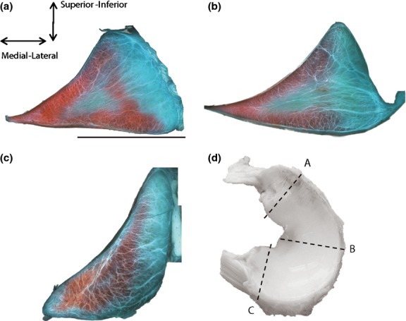
The collagenous structure of the knee menisci is integral to the mechanical integrity of the tissue and the knee joint. The tie-fibre structure of the tissue has largely been neglected, despite previous studies demonstrating its correlation with radial stiffness. This study has evaluated the structure of the tie-fibres of bovine menisci using 2D and 3D microscopy techniques. Standard collagen and proteoglycan (PG) staining and 2D light microscopy techniques were conducted. For the first time, the collagenous structure of the menisci was evaluated using 3D, second harmonic generation (SHG) microscopy. This technique facilitated the imaging of collagen structure in thick sections (50-100 μm). Imaging identified that tie-fibres of the menisci arborize from the outer margin of the meniscus toward the inner tip. This arborization is associated with the structural arrangement of the circumferential fibres. SHG microscopy has definitively demonstrated the 3D organization of tie-fibres in both sheets and bundles. The hierarchy of the structure is related to the organization of circumferential fascicles. Large tie-fibre sheets bifurcate into smaller sheets to surround circumferential fascicles of decreasing size. The tie-fibres emanate from the lamellar layer that appears to surround the entire meniscus. At the tibial and femoral surfaces these tie-fibre sheets branch perpendicularly into the meniscal body. The relationship between tie-fibres and blood vessels in the menisci was also observed in this study. Tie-fibre sheets surround the blood vessels and an associated PG-rich region. This subunit of the menisci has not previously been described. The size of tie-fibre sheets surrounding the vessels appeared to be associated with the size of blood vessel. These structural findings have implications in understanding the mechanics of the menisci. Further, refinement of the complex structure of the tie-fibres is important in understanding the consequences of injury and disease in the menisci. The framework of meniscus architecture also defines benchmarks for the development of tissue-engineered replacements in the future.
Keywords: blood vessels; meniscus; second harmonic generation microscopy; structure; tie-fibres.
© 2014 Anatomical Society.
Figures


References
-
- Bélisle J, Zigras T, Costantino S, et al. Second harmonic generation microscopy to investigate collagen configuration: a pericarditis case study. Cardiovasc Pathol. 2010;19:e125–e128. - PubMed
-
- Benninghoff A. Form und bau der Geleknorpel in ihren Bezeihungen zur Funktion. Z Zellforsch Mikrosk. 1925;2:783–825.
-
- Bullough PG, Munuera L, Murphy J, et al. The strength of the menisci of the knee as it relates to their fine structure. J Bone Joint Surg Br. 1970;52:564–567. - PubMed
-
- Campagnola PJ, Loew LM. Second-harmonic imaging microscopy for visualizing biomolecular arrays in cells, tissues and organisms. Nat Biotechnol. 2003;21:1356–1360. - PubMed
Publication types
MeSH terms
Substances
LinkOut - more resources
Full Text Sources
Other Literature Sources
Miscellaneous
