. 2014 Mar;16(2):92-100.
doi: 10.1111/ede.12067.

The evolution of the vertebrate cerebellum: absence of a proliferative external granule layer in a non-teleost ray-finned fish

Affiliations

The evolution of the vertebrate cerebellum: absence of a proliferative external granule layer in a non-teleost ray-finned fish

Thomas Butts et al. Evol Dev. 2014 Mar.

Abstract

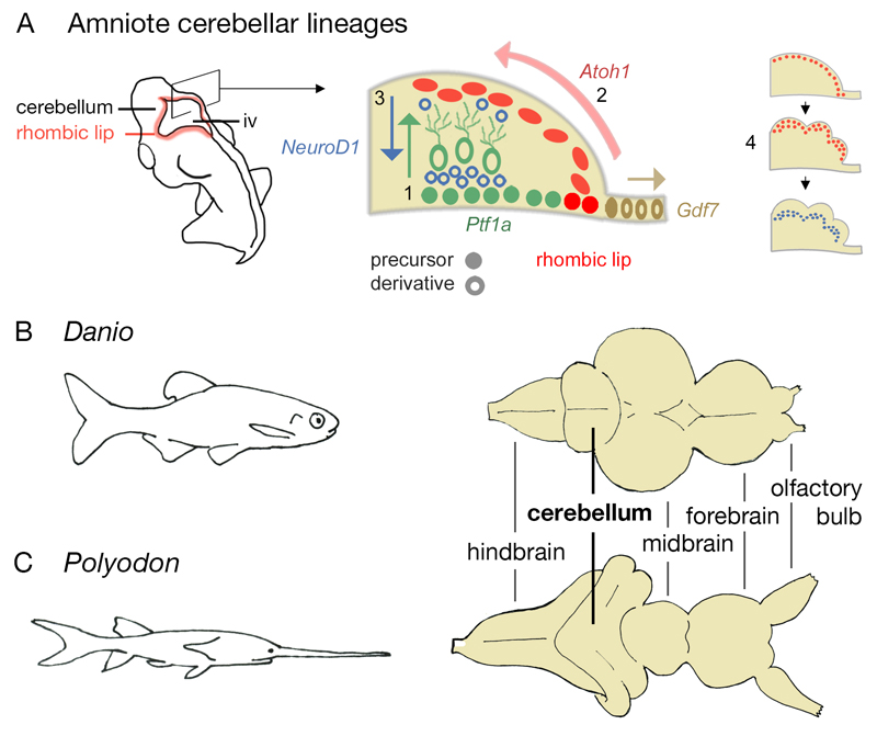
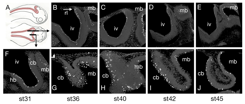
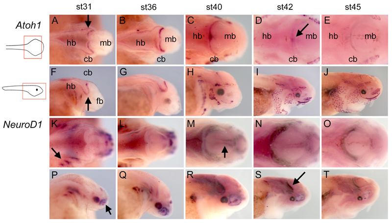
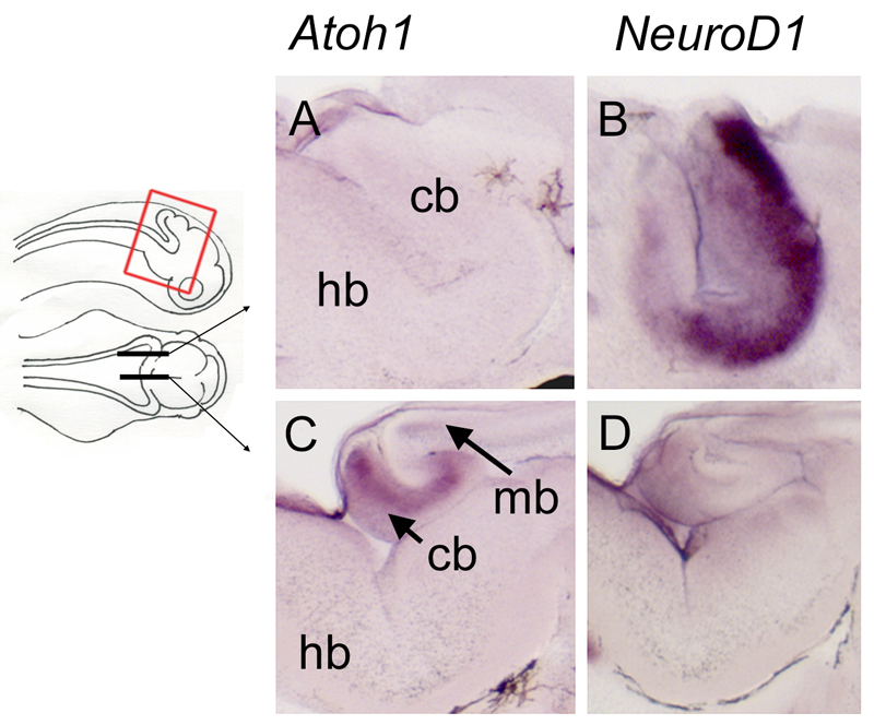
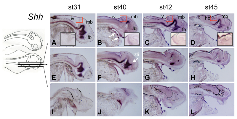
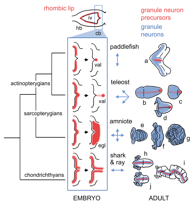
The cerebellum represents one of the most morphologically variable structures in the vertebrate brain. To shed light on its evolutionary history, we have examined the molecular anatomy and proliferation of the developing cerebellum of the North American paddlefish, Polyodon spathula. Absence of an external proliferative cerebellar layer and the restriction of Atonal1 expression to the rhombic lip and valvular primordium demonstrate that transit amplification in a cerebellar external germinal layer, a prominent feature of amniote cerebellum development, is absent in paddlefish. Furthermore, expression of Sonic hedgehog, which drives secondary proliferation in the mouse cerebellum, is absent from the paddlefish cerebellum. These data are consistent with what has been observed in zebrafish and suggest that the transit amplification seen in the amniote cerebellum was either lost very early in the ray-finned fish lineage or evolved in the lobe-finned fish lineage. We also suggest that the Atoh1-positive proliferative valvular primordium may represent a synapomorphy (shared derived character) of ray-finned fishes. The topology of valvular primordium development in paddlefish differs significantly from that of zebrafish and correlates with the adult cerebellar form. The distribution of proliferative granule cell precursors in different vertebrate taxa is thus the likely determining factor in cerebellar morphological diversity.


Figures

Figure 1
Figure 1. Cerebellar development in amniotes and adult cerebellar form in zebrafish and paddlefish.
Schematic diagram of an amniote embryo (A) showing the location of the rhombic lip and, in section, the distribution of precursors and their derivatives. Three major lineages give rise to Purkinje and other GABAergic neurons (Ptf1a, green), granule cells (Atoh1, red) and roof plate (Gdf7, brown). Purkinje cells migrate radially from the ventricular layer (1). Granule cells migrate tangentially to form a proliferative external germinal layer (2) and then, as NeuroD1-positive post-mitotic neurons, migrate radially to form an internal granule layer (3). Proliferation within the Atoh1-positive external germinal layer (4) leads to transverse foliation of the adult amniote cerebellum (see Fig.6e,f,g). The cerebellum of the zebrafish (Danio rerio) (B) differs from that of a paddlefish (Polyodon spathula) (C, normalised to the same scale as B): the latter displays a ribbon-like cerebellum with a convoluted and extended cerebellar rhombic lip. iv, fourth ventricle.
Figure 2
Figure 2. Proliferation in the paddlefish hindbrain.
Schematic diagram (A) of a paddlefish embryo in lateral and dorsal aspect showing the rhombic lip in red and the location of the parasagittal and sagittal sections. Immunostaining for phosphohistone H3 reveals that M-phase cells in the ventricular layer of the fourth ventricle (iv) progressively decline in number between stages 36 (B), 40 (C), 42 (D) and 45 (E), but continue to populate the midbrain (mb) ventricular surface. Sagittal sections at stages 31 (F), 36 (G), 40 (H), 42 (I) and 45(J) showing that proliferation is selectively maintained in the midline cerebellum (cb). Anterior is to the right in all panels. Abbreviations: cb, cerebellum; hb, hindbrain; iv, fourth ventricle; mb, midbrain; rl, rhombic lip.
Figure 3
Figure 3. Atoh1 and NeuroD1 expression in wholemount paddlefish embryos.
Whole mount in situ hybridisation for Atoh1 (A–J) and NeuroD1 (K–T) in paddlefish embryos between stages 31 – 45 in dorsal (A–E, K–O) and lateral (F–J, P–T) views. Arrows in A and F indicate the characteristic out-pocketing of the chondrostean cerebellar rhombic lip (Nieuwenhuys, ten Donkelaar, and Nicholson 1998). The arrow in D indicates the refinement of Atoh1 expression to the cerebellum midline at st42. Arrows in K and P, respectively, indicate NeuroD1 expression in the cranial ganglia and olfactory pit. The arrow in M indicates midbrain expression of NeuroD1, corresponding to the position of the mesencephalic trigeminal nucleus (arrow). The arrow in S indicates the position of cerebellar NeuroD1 expression. Abbreviations: cb, cerebellum; fb, forebrain; hb, hindbrain; iv, fourth ventricle; mb, midbrain; rl, rhombic lip.
Figure 4
Figure 4. Complementary expression of Atoh1 and NeuroD1 in the medial versus lateral rhombic lip at stage 42.
Sections through the rhombic lip of embryos after in situ hybridisation for Atoh1 and NeuroD1 in parasagittal (A,B) and sagittal (C,D) planes, as indicated on the schematic. In the lateral rhombic lip, Atoh1-positive progenitors are absent (A) but NeuroD1 is strongly expressed (B), consistent with previously reported distributions of granule neurons (Nieuwenhuys 1967). In the medial rhombic lip, i.e., the presumptive valvulus, Atoh1 is strongly expressed (C), while NeuroD1 is reduced (D). Abbreviations: cb, cerebellum; hb, hindbrain; mb, midbrain.
Figure 5
Figure 5. Sonic hedgehog is not expressed in the developing paddlefish cerebellum.
Sections through embryos from stages 31-45 after in situ hybridisation for Shh in sagittal (A-D) and parasagittal (E-L) planes, as indicated in the schematic. At all stages examined, Shh expression in the CNS is restricted to the ventral midline, with no expression in the cerebellum (red boxes, shown at higher power in insets). Shh expression in the CNS is lost further laterally (compare E-H with I-L). White arrows in B indicate prominent expression of Shh in tooth buds. The arrow in F indicates the zona limitans intrathalamica (zli), which identifies the mid-point of the diencephalon along the rostrocaudal axis. The dotted line in I demarcates the boundary of the ventral neural tube. Abbreviations: fb, forebrain; hb, hindbrain; iv, fourth ventricle; mb, midbrain; zli, zona limitans intrathalamica.
Figure 6
Figure 6. Hypothetical phylogeny of granule neuron proliferative zones in the cerebellum.
Hindbrain development in all vertebrate embryos can be characterised by a phase (top) in which an expanded fourth ventricle roof plate (iv) is bordered by an Atoh1-positive rhombic lip (red) in both the prospective cerebellum (cb) and the rest of the hindbrain (hb). Only the “upper” or cerebellar rhombic lip gives rise to granule cells in taxon-specific ways. We hypothesise that variation in the development of these proliferative zones (blue boxed schematics) constrains the geometry of the expansion of the granule cell layer (blue arrows, right) to produce mature granule cell distributions (blue) in adult, taxon-specific cerebellar morphologies (far right). In actinopterygians, granule cell proliferation becomes confined to a stem cell niche, the valvular primordium (val), which lies at the rostral pole of the cerebellum in teleosts (Kaslin et al. 2009) and borders the roof plate in the paddlefish. In amniotes, granule cell precursors migrate into a transient superficial external germinal layer (egl) and no precursors are retained in the adult cerebellum. In chondrichthyans, granule neuron progenitors are confined to the rhombic lip and on either side of the cerebellar midline (Chaplin, Tendeng, and Wingate 2010). Figures show hypothetical cell distributions in the cerebellar outlines (not shown to the same scale) based on model species for (a) paddlefish (a chondrostean ray-finned fish); (b-d) teleosts: b, remora (a perciform teleost), c, zebrafish (a cypriniform teleost), d, catfish (a siluriform teleost); (e-g) amniotes: e, bird, f, bat, g, giraffe; (h-j) chondrichthyans: h, dogfish (a shark), i, skate, j, stingray. Abbreviations: cb, cerebellum; egl, external germinal layer; hb, hindbrain; iv, fourth ventricle; val, valvular primordium.

References

    1. Abascal F, Zardoya R, Posada D. ProtTest: selection of best-fit models of protein evolution. Bioinformatics. 2005;21(9):2104–5. - PubMed
    1. Alder J, Cho NK, Hatten ME. Embryonic precursor cells from the rhombic lip are specified to a cerebellar granule neuron identity. Neuron. 1996;17(3):389–99. - PubMed
    1. Bemis WE, Grande L. Early development of the acitnopterygian head. 1. External development and staging of the paddlefish Polydon spatula. Journal of Morphology. 1992;213:47–83. - PubMed
    1. Ben-Arie N, Bellen HJ, Armstrong DL, McCall AE, Gordadze PR, Guo Q, Matzuk MM, Zoghbi HY. Math1 is essential for genesis of cerebellar granule neurons. Nature. 1997;390(6656):169–72. - PubMed
    1. Braitenberg V, Atwood RP. Morphological observations on the cerebellar cortex. J Comp Neurol. 1958;109:1–34. - PubMed

Publication types

Substances

LinkOut - more resources