. 2014 Apr 28;55(4):2452-9.
doi: 10.1167/iovs.13-13723.

A Pro23His mutation alters prenatal rod photoreceptor morphology in a transgenic swine model of retinitis pigmentosa

Affiliations

A Pro23His mutation alters prenatal rod photoreceptor morphology in a transgenic swine model of retinitis pigmentosa

Patrick A Scott et al. Invest Ophthalmol Vis Sci. .

Abstract

Purpose: Functional studies have detected deficits in retinal signaling in asymptomatic children from families with inherited autosomal dominant retinitis pigmentosa (RP). Whether retinal abnormalities are present earlier during gestation or shortly after birth in a subset of children with autosomal dominant RP is unknown and no appropriate animal RP model possessing visual function at birth has been available to examine this possibility. In a recently developed transgenic P23H (TgP23H) rhodopsin swine model of RP, we tracked changes in pre- and early postnatal retinal morphology, as well as early postnatal retinal function.

Methods: Domestic swine inseminated with semen from a TgP23H miniswine founder produced TgP23H hybrid and wild type (Wt) littermates. Outer retinal morphology was assessed at light and electron microscopic levels between embryonic (E) and postnatal (P) day E85 to P3. Retinal function was evaluated using the full field electroretinogram at P3.

Results: Embryonic TgP23H rod photoreceptors are malformed and their rhodopsin expression pattern is abnormal. Consistent with morphological abnormalities, rod-driven function is absent at P3. In contrast, TgP23H and Wt cone photoreceptor morphology (E85-P3) and cone-driven retinal function (P3) are similar.

Conclusions: Prenatal expression of mutant rhodopsin alters the normal morphological and functional development of rod photoreceptors in TgP23H swine embryos. Despite this significant change, cone photoreceptors are unaffected. Human infants with similarly aggressive RP might never have rod vision, although cone vision would be unaffected. Such aggressive forms of RP in preverbal children would require early intervention to delay or prevent functional blindness.

Keywords: electron microscopy; electroretinography; photoreceptor morphology; retinitis pigmentosa; swine.


Figures

Figure 1
Figure 1
Light microscopy and morphometric analysis of Wt and TgP23H swine outer retina (E85-P3). (A) Retinal micrographs taken 5 mm above optic disc in Wt versus TgP23H swine. Wt sections (left column) show the normal laminar arrangement of the retina during histogenesis. Retinae from TgP23H swine at E85 (right column) exhibit no change compared with Wt. TgP23H swine at E105 through P3 lack rod outer segments (ROS); CISs appear enlarged and their outer segments (black arrows) abut the RPE, and the ONL appears thinner at P0 and P3. Scale bar: 20 μm and applies to all panels. (B) Mean thickness (averaged across all locations and all eyes) of the ONL (E85–P3) in Wt versus TgP23H swine. P0 and P3 exhibit overall significant reduction in ONL thickness. (C) Mean thickness (averaged at each tested location) of the ONL along the vertical and horizontal meridia in Wt versus TgP23H. P0 and P3 TgP23H retinae show a central-to-peripheral pattern of thinning of the ONL. *P ≤ 0.05.
Figure 2
Figure 2
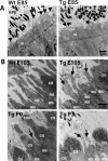
Ultrastructure of photoreceptor layer in Wt versus TgP23H swine. (A) TEM image of photoreceptor layer in Wt versus TgP23H E85. Photoreceptor IS and connecting-cilium (black arrowhead) appeared similar, and outer segments have not yet formed in Wt and Tg. Scale bar: 2 μm. (B) TEM image of photoreceptor layer in Wt versus TgP23H swine E105 through P3. E105 Wt, ROS/RIS, and COS/CIS exhibit normal morphology. E105 through P3 TgP23H, truncated ROS (black arrows) extend from RIS, COS appear normal or are lacking, and CIS appear enlarged. IS, inner segment; RIS, rod inner segment. Scale bar: 2 μm.
Figure 3
Figure 3
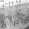
TEM image of outer retina TgP23H swine (P0). Cone outer segments (black arrows) abut the RPE and many CIS lack cone outer segments. External limiting membrane is intact (white arrows). Outer nuclear layer shows cone nuclei in the outermost row and stacks of rod nuclei (RN1–RN4), with degenerating rod nuclei (black arrowhead). Only cone pedicles (CP) can be seen in the outer plexiform layer. Scale bar: 10 μm.
Figure 4
Figure 4
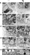
TEM image of outer plexiform layer Wt versus TgP23H swine (E85–P3). (A) E85, no identifiable photoreceptor axons or synaptic terminals in the OPL. (B) E105 Wt, axons extending from RN into OPL, as well as a few ribbons (black arrowheads), while TgP23H show no axons or ribbons. (C) P0 and (D) P3 Wt retinae show rod spherules (white arrows) and CP with synapses and triads in the OPL. E105 through P3 TgP23H retinae show axonal retraction, no spherules or triads, and few ribbon synapses (black arrows), but cone photoreceptor axons and pedicles appear normal. (E) Magnification of boxed in region in P0 showing Wt showing normal triadic profiles (white arrows) in Wt and their absence in TgP23H (black arrow). HZCN, horizontal cell nucleus. Scale bar: 2 μm and applies to all panels.
Figure 5
Figure 5
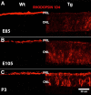
Immunolabeling with anti-Rho 1D4 (rhodopsin) antibody Wt versus TgP23H (E85–P3). (AC) Intense staining with anti-Rho 1D4 is restricted to the PRL in Wt retinae (left column). Anti-Rho 1D4 is delocalized to the ONL in TgP23H (right column). Scale bar: 20 μm and applies to all panels. Figures from P0 are not shown but are the same as P3.
Figure 6
Figure 6
Representative ffERG recordings for rod, cone, and 30-Hz flicker in Wt and TgP23H Swine (P3). (A) The rod waveform and histogram show a striking difference, with a nearly extinguished response in the TgP23H, while the Wt has a clearly discernible response. In contrast, the representative cone traces and histograms show how the morphology of the components of the cone and cone flicker waveforms at P3 ([B, C], respectively) are very similar between the TgP23H and Wt.

References

    1. Milam AH, Li Z-Y, Fariss RN. Histopathology of the human retina in retinitis pigmentosa. Prog Ret Eye Res. 1998; 17: 175–205 - PubMed
    1. Haim M. Prevalence of retinitis pigmentosa and allied disorders in Denmark. Acta Ophthalmol. 1992; 70: 615–624 - PubMed
    1. Weleber RG. Retinitis Pigmentosa and Allied Disorders. 2nd ed. St. Louis: Mosby; 1994: 335–466
    1. Berson EL, Gouras P, Gunkel RD. Rod response in retinitis pigmentosa, dominantly inherited. Arch Ophthalmol. 1968; 80: 58–67 - PubMed
    1. Berson EL, Simonoff EA. Dominant retinitis pigmentosa with reduced penetrance. Further studies with the electroretinogram. Arch Ophthalmol. 1979; 97: 1286–1291 - PubMed

Publication types

MeSH terms