. 2014 Mar 11;9(3):e91427.
doi: 10.1371/journal.pone.0091427. eCollection 2014.

Blood brain barrier and neuroinflammation are critical targets of IGF-1-mediated neuroprotection in stroke for middle-aged female rats

Affiliations

Blood brain barrier and neuroinflammation are critical targets of IGF-1-mediated neuroprotection in stroke for middle-aged female rats

Shameena Bake et al. PLoS One. .

Abstract

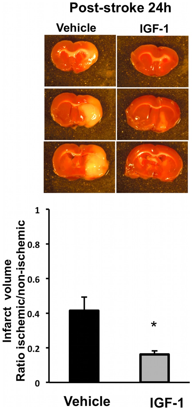
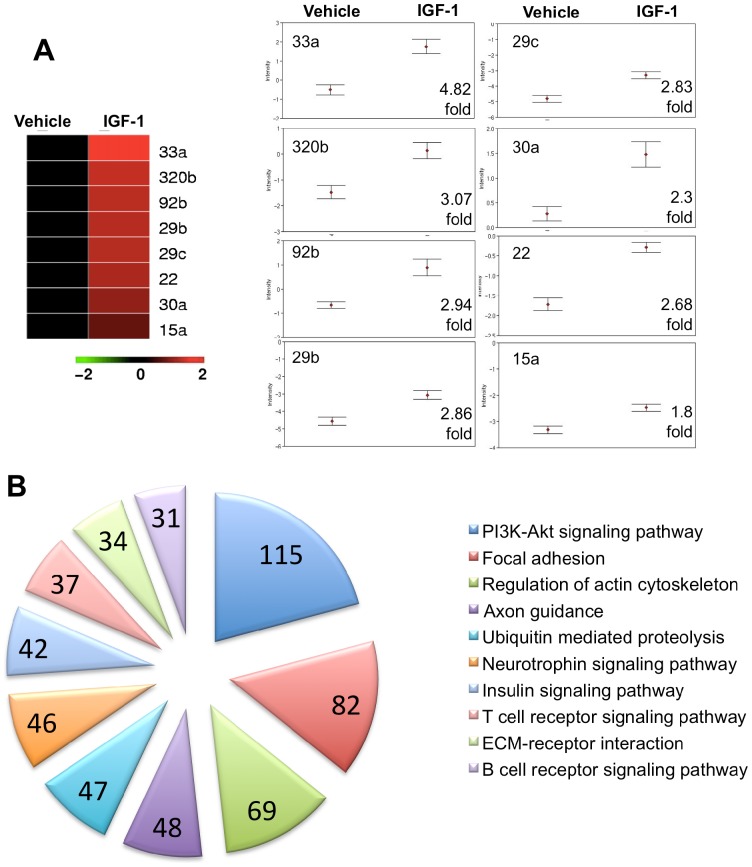
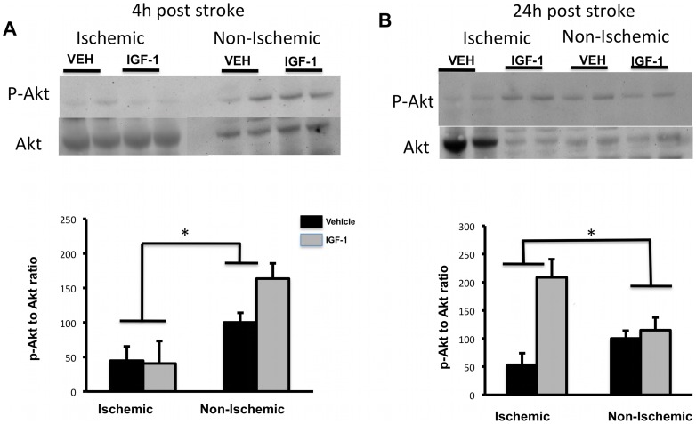
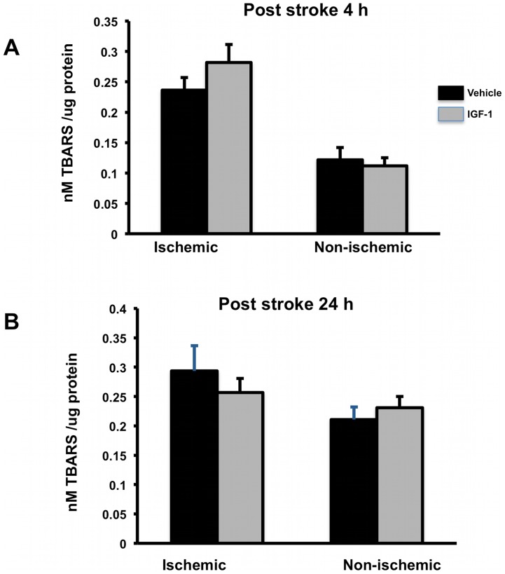
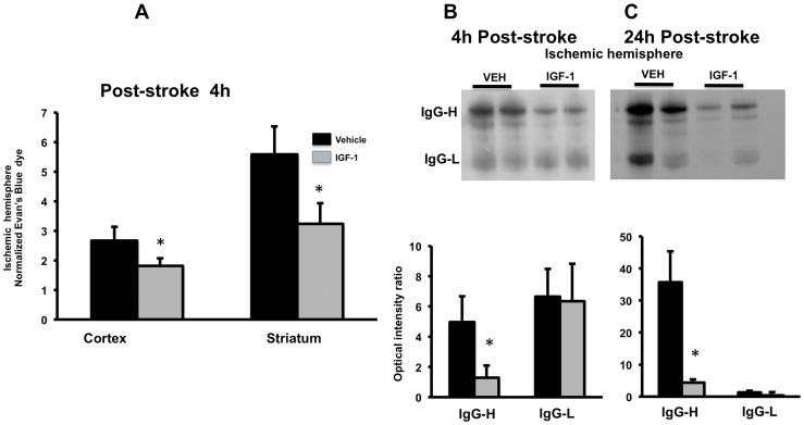
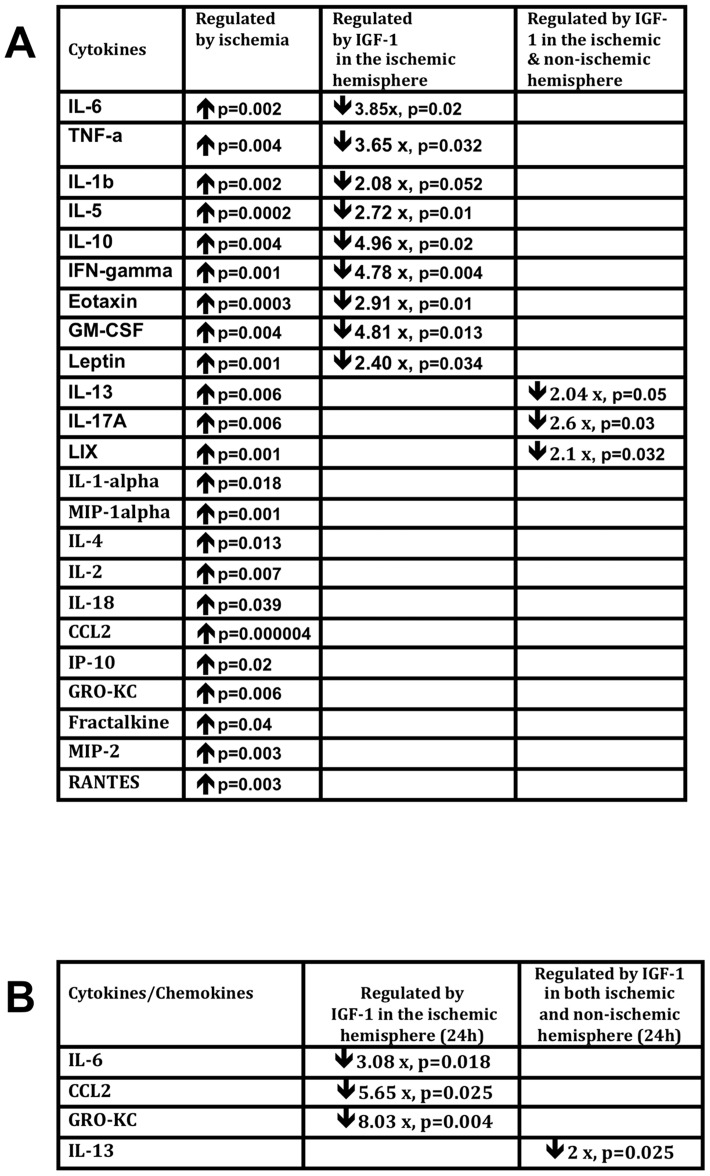
Ischemia-induced cerebral infarction is more severe in older animals as compared to younger animals, and is associated with reduced availability of insulin-like growth factor (IGF)-1. This study determined the effect of post-stroke IGF-1 treatment, and used microRNA profiling to identify mechanisms underlying IGF-1's neuroprotective actions. Post-stroke ICV administration of IGF-1 to middle-aged female rats reduced infarct volume by 39% when measured 24h later. MicroRNA analyses of ischemic tissue collected at the early post-stroke phase (4h) indicated that 8 out of 168 disease-related miRNA were significantly downregulated by IGF-1. KEGG pathway analysis implicated these miRNA in PI3K-Akt signaling, cell adhesion/ECM receptor pathways and T-and B-cell signaling. Specific components of these pathways were subsequently analyzed in vehicle and IGF-1 treated middle-aged females. Phospho-Akt was reduced by ischemia at 4h, but elevated by IGF-1 treatment at 24h. IGF-1 induced Akt activation was preceded by a reduction of blood brain barrier permeability at 4h post-stroke and global suppression of cytokines including IL-6, IL-10 and TNF-α. A subset of these cytokines including IL-6 was also suppressed by IGF-1 at 24h post-stroke. These data are the first to show that the temporal and mechanistic components of post-stroke IGF-1 treatment in older animals, and that cellular components of the blood brain barrier may serve as critical targets of IGF-1 in the aging brain.


Conflict of interest statement

Competing Interests: The authors have declared that no competing interests exist.

Figures

Figure 1
Figure 1. Effect of IGF-1 on infarct volume:
MCAo for 90 min followed by reperfusion resulted in a cortical-striatal infarction as seen in TTC-stained coronal sections. Quantitative analysis of infarct volume, expressed as a ratio to the non-ischemic hemisphere, shows that post-stroke IGF-1 treatment resulted in a 39% decrease in infarct size compared to control after 24 h. Bars represent mean ± SEM. N =  6–8 in each group. *: p <0.05.
Figure 2
Figure 2. MiRNA regulation by IGF-1 in the ischemic brain at 4h post stroke.
MiRNAs that were significantly regulated (adjusted for FDR) are graphically represented in a heat map format. Within the heat map, the vehicle-treated animals were used as ‘controls’ and are represented in black, while IGF-1 treated animals are depicted in relation to vehicle controls (columns). All miRNA in this cohort were significantly downregulated by IGF-1 (shown in red) in comparison vehicle controls (rows). (B) Group differences in expression patterns of IGF-1 regulated miRNA are shown graphically, with the ‘fold’ regulation indicated at the bottom right corner. Data (mean+SEM) are expressed are expressed as ΔCT, where an increased value indicates decreased miRNA expression. N  =  6/group p<0.05. (C) MiRNAs that were significantly regulated by IGF-1 treatment were subject to in silico analysis (DIANA/miRPATH, see methods). Predicted gene targets for each miRNA were identified. The top 10 KEGG pathways represented by these predicted targets are shown as a pie chart. Each slice represents the number of predicted target genes in the pathway, indicated within the slice. Each pathway is color coded and labeled adjacent to the chart. The top 10 pathways were selected based on the smallest “p” value and the largest number of target genes in the pathway.
Figure 3
Figure 3. Phospho-Akt and Akt expression in post-ischemic brain.
pAkt and pan-Akt levels were analyzed by Western blot (top panels). pAkt expression, normalized to pan-Akt, is shown in the histograms (lower panels). (A) p-Akt levels were reduced in both control and IGF-1-treated groups in the ischemic hemisphere at 4h, as compared to the non-ischemic hemisphere (main effect of ischemia; p<0.05). (B) At 24h, p-Akt expression was significantly increased in the ischemic hemisphere of the IGF-1-treated group as compared to the vehicle-treated group (interaction effect of ischemia and IGF-1). Bars represent mean ± SEM. n =  6 in each group. *: p <0.05.
Figure 4
Figure 4. Phospho-ERK and pan ERK expression in post-ischemic brain.
pERK and pan ERK were analyzed by Western blot (top panels). pERK expression, normalized to ERK, are shown in the histograms (lower panels). (A) p-ERK1 level increased in the ischemic hemisphere at 4h, as compared to the non-ischemic hemisphere (main effect of hemisphere, p<0.05). (B) At 24h, both p-ERK1 & 2 were significantly increased in the ischemic hemisphere (main effect of hemisphere; p<0.05). IGF-1 did not affect pERK expression at either time point. Bars represent mean ± SEM. n =  6 in each group. *: p <0.05.
Figure 5
Figure 5. Expression of TBARS:
(A) TBARS levels were increased with ischemia-reperfusion at 4h in both control and IGF-1 group compared to non-ischemic hemisphere. IGF-1 had no effect on TBARS levels. (B) At 24h post stroke, TBARS were similar in the ischemic and non-ischemic hemisphere and were not altered by IGF-1 treatment. Bars represent mean ± SEM. n =  6–8 in each group. *: p<0.05.
Figure 6
Figure 6. Effect of IGF-1 on ischemia-reperfusion induced blood brain barrier permeability.
(A). Evans blue dye extravasation: IGF-1 treatment significantly reduced ischemia-induced increase in blood brain barrier permeability at 4h as indicated by decreased Evan’s blue dye in both cortex and striatum. Concentration of Evan’s blue is expressed as ratio of dye accumulation in the ischemic hemisphere to the non-ischemic hemisphere. Bars represent mean ± SEM. n =  6–8 in each group. *: p <0.05. (B) IgG expression: Western blot analysis of light and heavy chain IgG expression indicated that IGF-1 treatment reduced IgG-heavy chain expression at both 4h and 24h post stroke.
Figure 7
Figure 7. IGF-1 mediated regulation of inflammatory cytokines in post-ischemic brain.
(A). Multiplex analysis of brain tissue lysates from the ischemic and non-ischemic hemisphere at 4h post stroke. The summary table shows each cytokine,and its regulation by ischemia or by IGF-1. Almost all cytokines were upregulated by ischemia. IGF-1 decreased the expression of approximately half of the cytokines on this panel (column 3; indicated by downward arrows and fold change), in the ischemic hemisphere and a few in both hemispheres (last column). (B) At 24 h, IL-6 and IL-13 continued to be suppressed in the IGF-1-treated group, as well as the chemokines GRO-KC and CCL2. n =  6-8 in each group; p<0.05 in each case.

References

    1. Faber JE, Zhang H, Lassance-Soares RM, Prabhakar P, Najafi AH, et al. (2011) Aging causes collateral rarefaction and increased severity of ischemic injury in multiple tissues. Arterioscler Thromb Vasc Biol 31: 1748–1756. - PMC - PubMed
    1. Towfighi A, Saver JL, Engelhardt R, Ovbiagele B (2007) A midlife stroke surge among women in the United States. Neurology 69: 1898–1904. - PubMed
    1. Rosamond W, Flegal K, Furie K, Go A, Greenlund K, et al. (2008) Heart disease and stroke statistics—2008 update: a report from the American Heart Association Statistics Committee and Stroke Statistics Subcommittee. Circulation 117: e25–146. - PubMed
    1. Turtzo LC, McCullough LD (2008) Sex differences in stroke. Cerebrovasc Dis 26: 462–474. - PMC - PubMed
    1. Petrea RE, Beiser AS, Seshadri S, Kelly-Hayes M, Kase CS, et al. (2009) Gender differences in stroke incidence and poststroke disability in the Framingham heart study. Stroke 40: 1032–1037. - PMC - PubMed

Publication types

MeSH terms