

Treadmill exercise induced functional recovery after peripheral nerve repair is associated with increased levels of neurotrophic factors
- PMID: 24618564
- PMCID: PMC3949693
- DOI: 10.1371/journal.pone.0090245
Treadmill exercise induced functional recovery after peripheral nerve repair is associated with increased levels of neurotrophic factors
Abstract
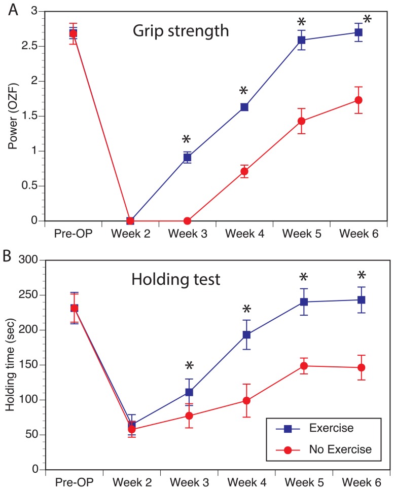
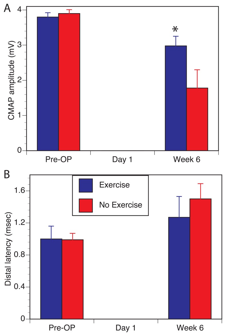
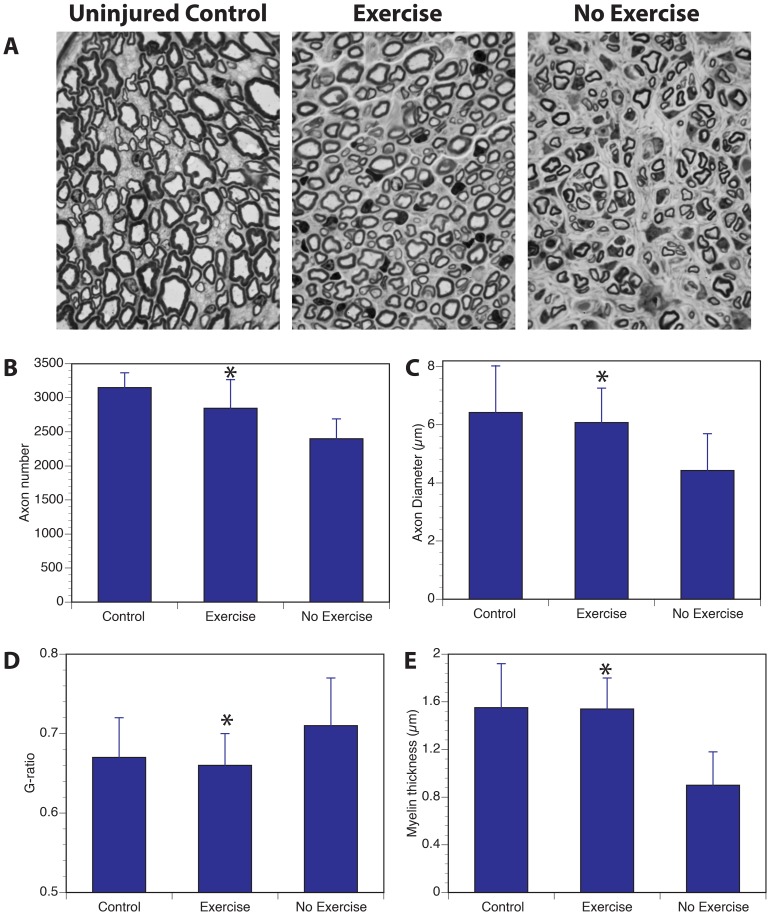
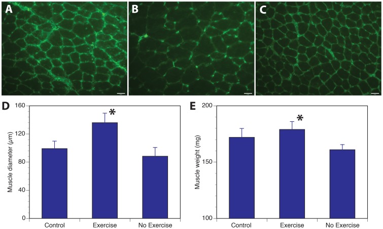
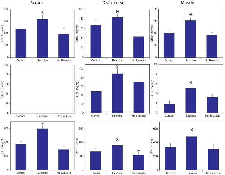
Benefits of exercise on nerve regeneration and functional recovery have been reported in both central and peripheral nervous system disease models. However, underlying molecular mechanisms of enhanced regeneration and improved functional outcomes are less understood. We used a peripheral nerve regeneration model that has a good correlation between functional outcomes and number of motor axons that regenerate to evaluate the impact of treadmill exercise. In this model, the median nerve was transected and repaired while the ulnar nerve was transected and prevented from regeneration. Daily treadmill exercise resulted in faster recovery of the forelimb grip function as evaluated by grip power and inverted holding test. Daily exercise also resulted in better regeneration as evaluated by recovery of compound motor action potentials, higher number of axons in the median nerve and larger myofiber size in target muscles. Furthermore, these observations correlated with higher levels of neurotrophic factors, glial derived neurotrophic factor (GDNF), brain derived neurotrophic factor (BDNF) and insulin-like growth factor-1 (IGF-1), in serum, nerve and muscle suggesting that increase in muscle derived neurotrophic factors may be responsible for improved regeneration.
Conflict of interest statement
Figures
References
-
- Mattson MP (2000) Neuroprotective signaling and the aging brain: take away my food and let me run. Brain Res 886: 47–53. - PubMed
-
- Haskell WL, Lee IM, Pate RR, Powell KE, Blair SN, et al. (2007) Physical activity and public health: updated recommendation for adults from the American College of Sports Medicine and the American Heart Association. Med Sci Sports Exerc 39: 1423–1434. - PubMed
-
- Edgerton VR, Tillakaratne NJ, Bigbee AJ, de Leon RD, Roy RR (2004) Plasticity of the spinal neural circuitry after injury. Annu Rev Neurosci 27: 145–167. - PubMed
-
- Hutchinson KJ, Gomez-Pinilla F, Crowe MJ, Ying Z, Basso DM (2004) Three exercise paradigms differentially improve sensory recovery after spinal cord contusion in rats. Brain 127: 1403–1414. - PubMed
Publication types
MeSH terms
Substances
LinkOut - more resources
Full Text Sources
Other Literature Sources
Medical
Miscellaneous
