. 2014 Mar 11;9(3):e90857.
doi: 10.1371/journal.pone.0090857. eCollection 2014.

A generic method for design of oligomer-specific antibodies

Affiliations

A generic method for design of oligomer-specific antibodies

Kristoffer Brännström et al. PLoS One. .

Abstract

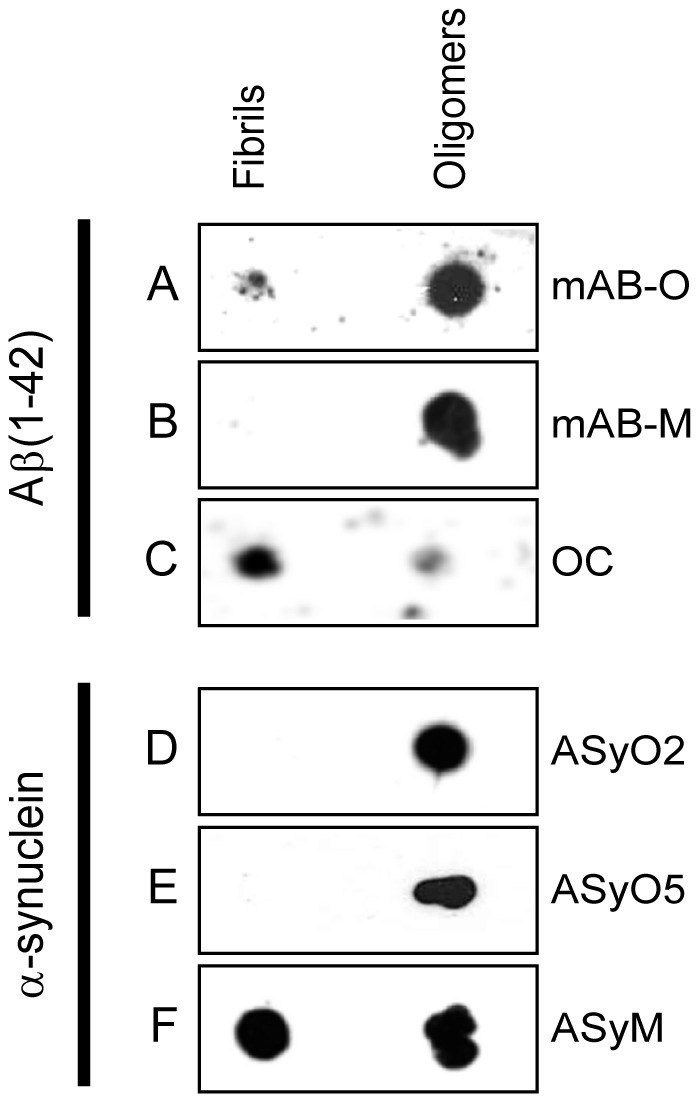
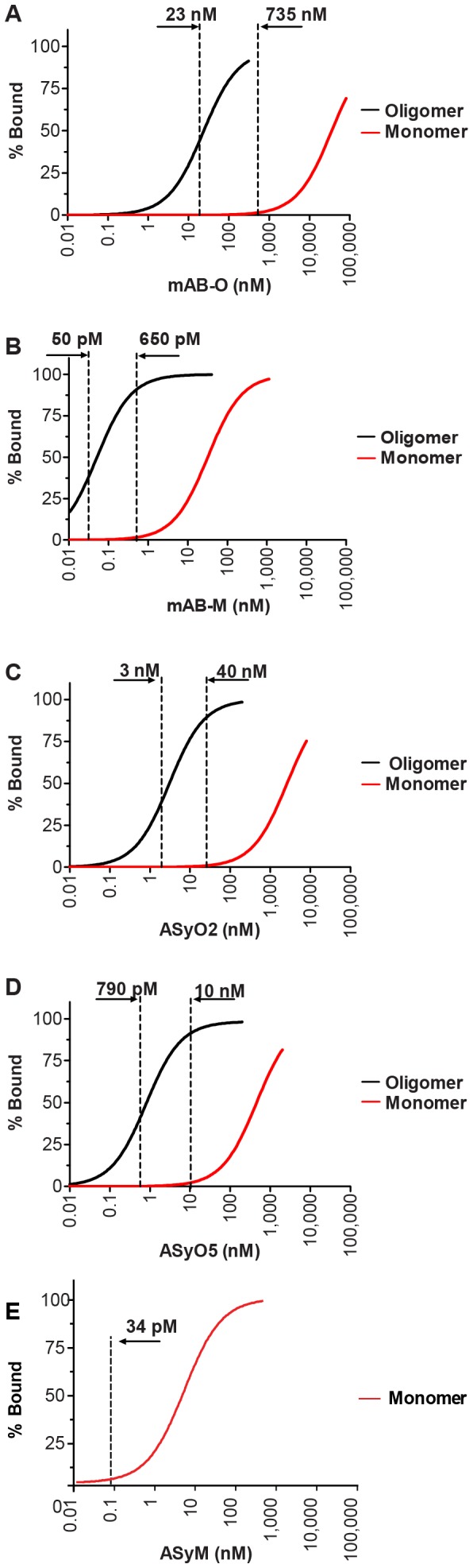
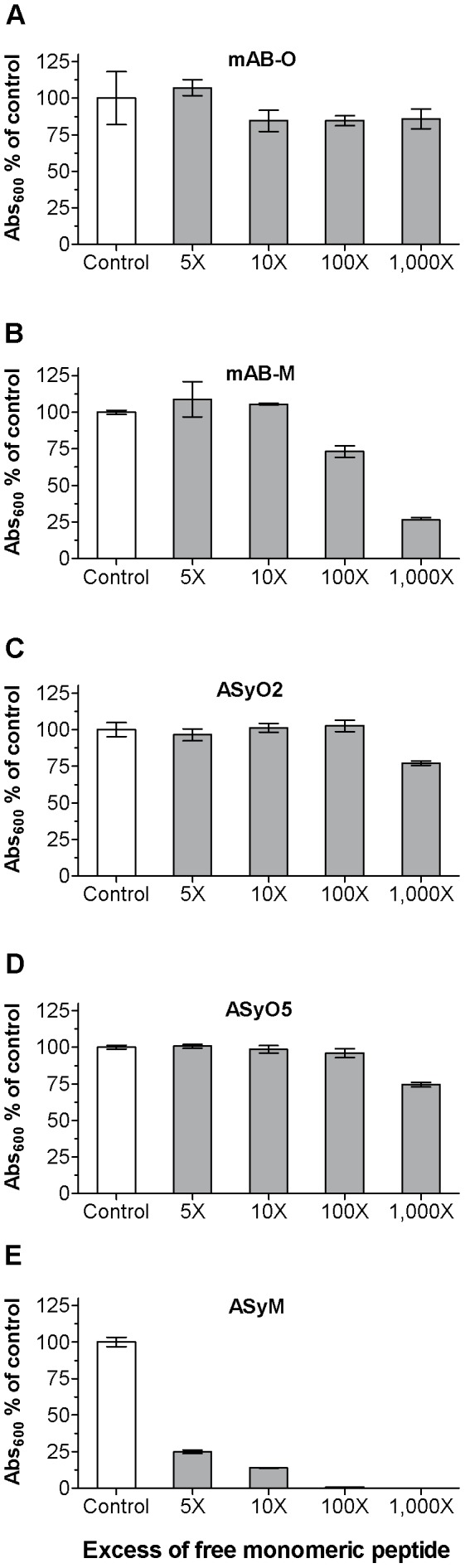
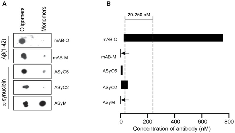
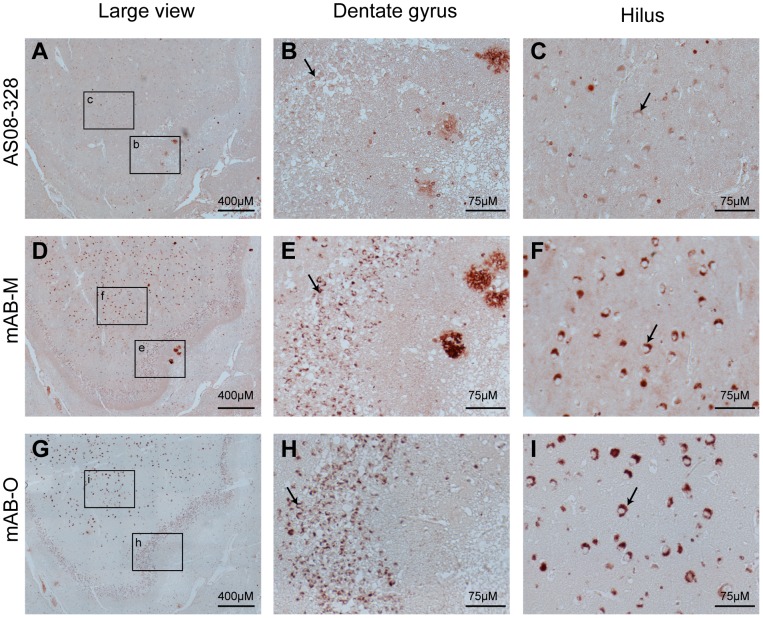
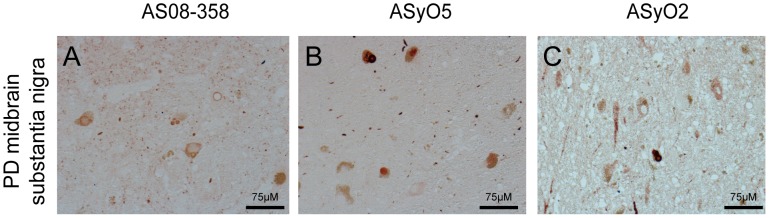
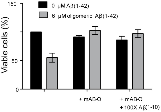
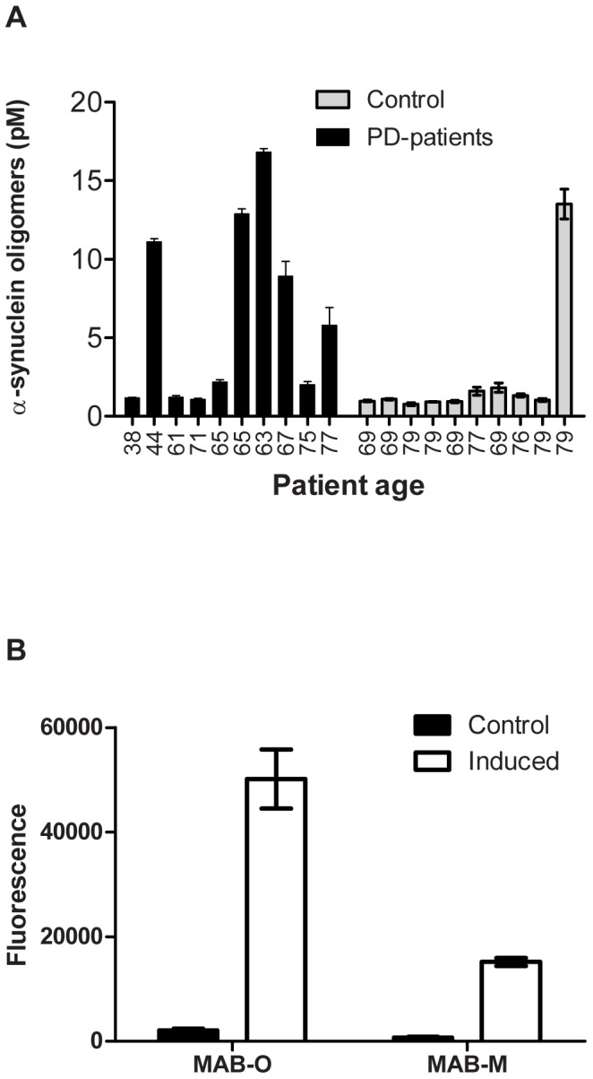
Antibodies that preferentially and specifically target pathological oligomeric protein and peptide assemblies, as opposed to their monomeric and amyloid counterparts, provide therapeutic and diagnostic opportunities for protein misfolding diseases. Unfortunately, the molecular properties associated with oligomer-specific antibodies are not well understood, and this limits targeted design and development. We present here a generic method that enables the design and optimisation of oligomer-specific antibodies. The method takes a two-step approach where discrimination between oligomers and fibrils is first accomplished through identification of cryptic epitopes exclusively buried within the structure of the fibrillar form. The second step discriminates between monomers and oligomers based on differences in avidity. We show here that a simple divalent mode of interaction, as within e.g. the IgG isotype, can increase the binding strength of the antibody up to 1500 times compared to its monovalent counterpart. We expose how the ability to bind oligomers is affected by the monovalent affinity and the turnover rate of the binding and, importantly, also how oligomer specificity is only valid within a specific concentration range. We provide an example of the method by creating and characterising a spectrum of different monoclonal antibodies against both the Aβ peptide and α-synuclein that are associated with Alzheimer's and Parkinson's diseases, respectively. The approach is however generic, does not require identification of oligomer-specific architectures, and is, in essence, applicable to all polypeptides that form oligomeric and fibrillar assemblies.


Conflict of interest statement

Competing Interests: The authors have declared that no competing interests exist.

Figures

Figure 1
Figure 1. Dot blot enables probing for structural differences between fibrils and oligomers.
An equal amount of fibrils and oligomers from either Aβ(1–42) or α-synuclein, as indicated in the figure, were applied to a nitrocellulose membrane. Binding specificity of (A) mAB-O, (B) mAB-M, (C) Aβ fibril-specific OC antibody,, (D) ASyO2, (E) ASyO5, and (F) ASyM.
Figure 2
Figure 2. Monovalent and divalent saturation level as a function of concentration.
The level of antibody saturation upon binding to oligomers is shown as a black line and binding to monomers is shown as a red line. The vertical dotted lines indicate the concentrations where the saturation level of binding to the oligomer is above 50% while the monovalent saturation level remains below 2%. (A) mAB-O, (B) mAB-M, (C) ASyO2, (D) ASyO5, (E) ASyM.
Figure 3
Figure 3. The rate of turnover controls antibody efficacy.
Competition ELISA setup showing the efficacy of the antibodies to detect oligomers in the presence of increasing concentrations of monomers. (A) mAB-O. (B) mAB-M. (C) ASyO2. (D) ASyO5. (E) ASyM. The kinetics of binding for each antibody are given in Table 1.
Figure 4
Figure 4. Therapeutic antibody concentrations require very low monovalent affinities to avoid reactivity to monomers.
(A)Non-logarithmic illustration of the antibody concentration range where more than 50% of the oligomers are bound while the saturation level to the monomer is below 2%. Note in particular the dramatic difference between mAB-O and mAB-M that both qualify as being oligomer specific according to the widely accepted dot blot technique. The bars corresponding to mAB-M as well as ASyM are indicated with arrows to highlight their appearance. For comparison, the most commonly used range of therapeutic antibody concentrations for AD (20 µM to 250 µM) is indicated in the figure (grey striped vertical bars). (B) Dot blot analysis where equal amounts of Aβ1–42 and α-synuclein oligomers and monomers has been applied on a membrane and probed with mAB-O, mAB-M, ASyO5, ASyO2, and ASyM as indicated in the figure.
Figure 5
Figure 5. IHC of human hippocampus from an AD-affected individual.
IHC was used to illustrate binding specificity and structural preferences of mAB-O and mAB-M upon binding to ex vivo material. Hippocampus sections were from a post-mortem AD-affected human brain (A–C) The binding pattern from a polyclonal rabbit anti-Aβ antibody illustrating both plaques as well as an intracellular form of Aβ that is abundant in the hilar neurons. (D–F) The staining pattern of mAB-M in which Aβ plaques near the dentate gyrus (E) are readily stained. (G–I). The binding pattern of mAB-O illustrates an inability to stain Aβ plaques and indicates an alternative structural preference compared to mAB-M. A strong binding to the intracellular form of Aβ is observed in the hilus area. The granular cells of the dentate gyrus (B, E, and H) and the neurons of hilus (C, F, and I) are indicated with arrows and represent the highe magnification images of respective selected areas in A, D and G.
Figure 6
Figure 6. Immunohistochemistry of human midbrain from a PD-affected individual.
To probe the specificity and structural preference of ASyO2 towards ex vivo α-synuclein assemblies, the mesencephalon (midbrain) of the brain of a human PD patient was analysed. The images represent the substantia nigra area of the midbrain. (A) Polyclonal anti α-synuclein. (B) ASyO5. (C) ASyO2.
Figure 7
Figure 7. Binding to biologically active oligomers of Aβ1–42.
Isolated Aβ1–42 oligomers exert a cytotoxic effect on SH-SY5Y cells, and the attenuating effect of the oligomer-specific mAB-O antibody was evaluated. Cells exposed to Aβ1–42 oligomers are shown with grey bars while controls are depicted in black. Addition of mAB-O in a 1∶5 molar ratio to Aβ1–42 fully attenuates the effect. Addition of Aβ1–10 monomer at a 100-fold molar excess does not interfere with the attenuating effect.
Figure 8
Figure 8. Detection of ex vivo-derived oligomeric assemblies.
(A) CSF samples from 10 PD patients having a clinical stage of disease around 2 according to the Hoehn and Yahr scale and 10 age-matched controls were probed for the presence of α-synuclein oligomers using a homopaired sandwich ELISA based on the ASyO2 antibody. The age of each individual is given below the corresponding bar. The concentration of α-synuclein oligomers was determined using a standard curve of in vitro α-synuclein oligomers based on their monomeric protein concentration. (B) A sandwich ELISA setup using mAB-O and mAB-M to evaluate binding to Aβ oligomers derived from the well-established and previously described 7PA2 cells .

References

    1. Sipe JD, Benson MD, Buxbaum JN, Ikeda S, Merlini G, et al. (2012) Amyloid fibril protein nomenclature: 2012 recommendations from the Nomenclature Committee of the International Society of Amyloidosis. Amyloid 19: 167–170. - PubMed
    1. Larson ME, Lesne SE (2012) Soluble Abeta oligomer production and toxicity. J Neurochem 120 Suppl 1: 125–139. - PMC - PubMed
    1. Lasagna-Reeves CA, Castillo-Carranza DL, Guerrero-Muoz MJ, Jackson GR, Kayed R (2010) Preparation and characterization of neurotoxic tau oligomers. Biochemistry 49: 10039–10041. - PubMed
    1. Lambert MP, Barlow AK, Chromy BA, Edwards C, Freed R, et al. (1998) Diffusible, nonfibrillar ligands derived from Abeta1–42 are potent central nervous system neurotoxins. Proc Natl Acad Sci U S A 95: 6448–6453. - PMC - PubMed
    1. Walsh DM, Klyubin I, Fadeeva JV, Cullen WK, Anwyl R, et al. (2002) Naturally secreted oligomers of amyloid beta protein potently inhibit hippocampal long-term potentiation in vivo. Nature 416: 535–539. - PubMed

Publication types

MeSH terms