Review
. 2014 Mar 1;5(2):144-63.
doi: 10.3945/an.113.005181.

From cholesterogenesis to steroidogenesis: role of riboflavin and flavoenzymes in the biosynthesis of vitamin D

Affiliations
Review

From cholesterogenesis to steroidogenesis: role of riboflavin and flavoenzymes in the biosynthesis of vitamin D

John T Pinto et al. Adv Nutr. .

Abstract

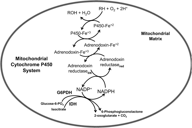
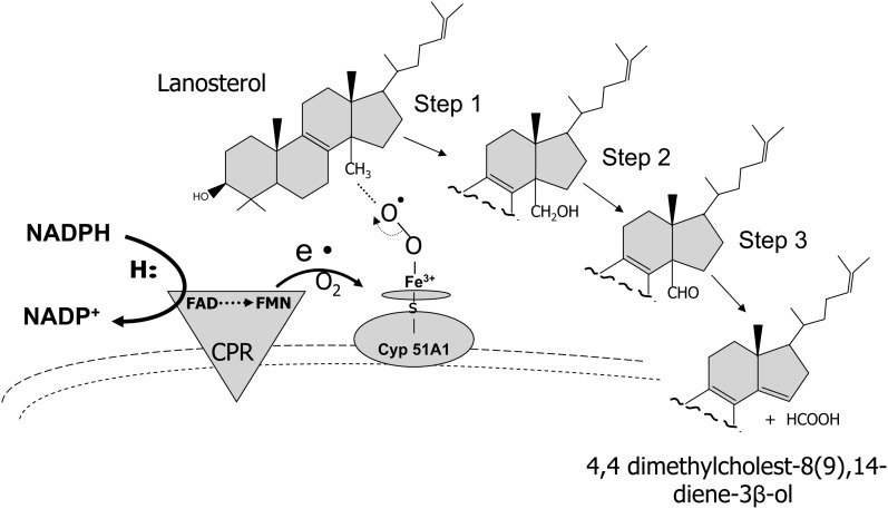
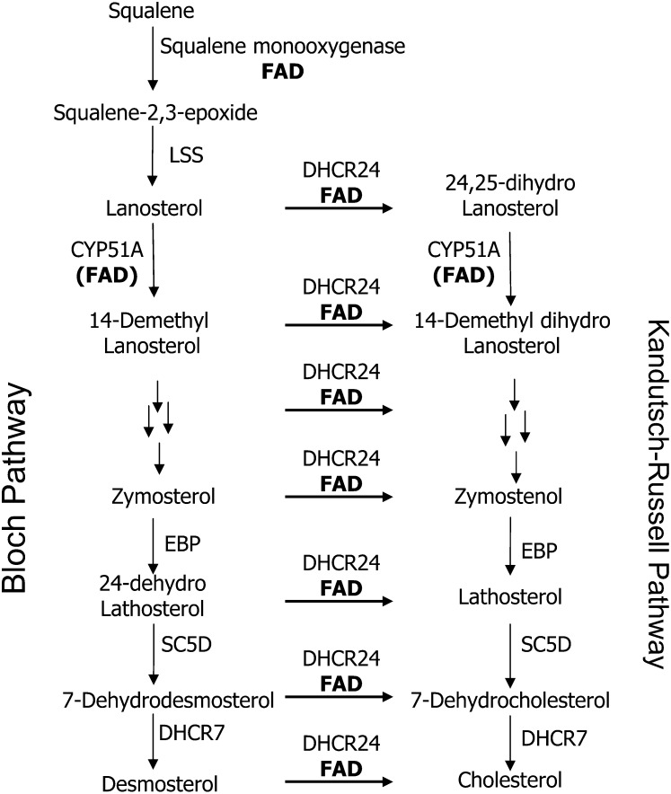
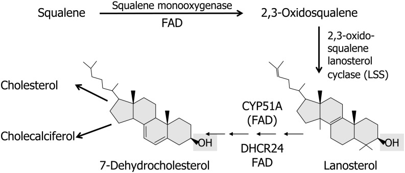
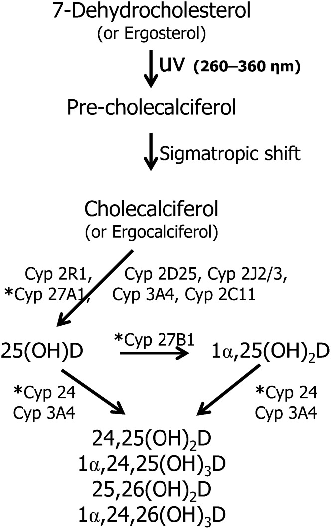
Flavin-dependent monooxygenases and oxidoreductases are located at critical branch points in the biosynthesis and metabolism of cholesterol and vitamin D. These flavoproteins function as obligatory intermediates that accept 2 electrons from NAD(P)H with subsequent 1-electron transfers to a variety of cytochrome P450 (CYP) heme proteins within the mitochondria matrix (type I) and the (microsomal) endoplasmic reticulum (type II). The mode of electron transfer in these systems differs slightly in the number and form of the flavin prosthetic moiety. In the type I mitochondrial system, FAD-adrenodoxin reductase interfaces with adrenodoxin before electron transfer to CYP heme proteins. In the microsomal type II system, a diflavin (FAD/FMN)-dependent cytochrome P450 oxidoreductase [NAD(P)H-cytochrome P450 reductase (CPR)] donates electrons to a multitude of heme oxygenases. Both flavoenzyme complexes exhibit a commonality of function with all CYP enzymes and are crucial for maintaining a balance of cholesterol and vitamin D metabolites. Deficits in riboflavin availability, imbalances in the intracellular ratio of FAD to FMN, and mutations that affect flavin binding domains and/or interactions with client proteins result in marked structural alterations within the skeletal and central nervous systems similar to those of disorders (inborn errors) in the biosynthetic pathways that lead to cholesterol, steroid hormones, and vitamin D and their metabolites. Studies of riboflavin deficiency during embryonic development demonstrate congenital malformations similar to those associated with genetic alterations of the flavoenzymes in these pathways. Overall, a deeper understanding of the role of riboflavin in these pathways may prove essential to targeted therapeutic designs aimed at cholesterol and vitamin D metabolism.


Conflict of interest statement

Author disclosures: J. T. Pinto and A. J. L. Cooper, no conflicts of interest.

Figures

FIGURE 1
FIGURE 1
Structures of flavin coenzymes. The isoalloxazine ring is shown relative to riboflavin and its nucleotide coenzymes, riboflavin 5′-phosphate (FMN) and FAD. The shaded area depicts the region of the isoalloxazine ring that is involved in the direct transfer of electrons from the flavoenzyme. Once transferred, electrons can migrate within the heterocyclic isoalloxazine ring.
FIGURE 2
FIGURE 2
Nucleophilic attack at the N5 or C4a position of the isoalloxazine ring with formation of a flavosemiquinone. Flavosemiquinones exist either in a neutral radical (FAD) or in an anionic radical (FAD•−) state. X and Y represent nucleophiles.
FIGURE 3
FIGURE 3
Mitochondrial CYP electron transfer system (type I). IDH and G6PDH generate NAD(P)H, which transfers a hydride ion (H:) through π-π interaction between the nicotinamide ring of NAD(P)H and the isoalloxazine moiety of FAD-adrenodoxin reductase. A single electron transfer to heme-containing adrenodoxin generates a ferro-heme moiety that interfaces with a variety of CYP heme enzymes. For example, a CYP enzyme (CYP27B1) catalyzes the oxidation of substrate RH (e.g, 25-hydroxyvitamin D) by adding 1 oxygen atom of molecular oxygen to the substrate (RH), producing a hydroxylated product ROH (e.g., 1α,25-dihydroxyvitamin D); the other atom of oxygen is reduced to water. CYP, cytochrome P450; IDH, isocitrate dehydrogenase; G6PDH, glucose-6-phosphate dehydrogenase; ox, oxidized form of adrenodoxin reductase; red, reduced form of adrenodoxin reductase.
FIGURE 4
FIGURE 4
Formation of squalene epoxides. Squalene monooxygenase catalyzes oxidation of squalene to form 2,3-oxidosqualene. This mixed-function oxidase requires NAD(P)H as the reductant and molecular O2 as oxidant. One oxygen atom of molecular oxygen is incorporated into the substrate (as the epoxide), and the other oxygen atom is reduced to water. An additional side reaction in the presence of excess FAD (bold letters) can occur with 2,3-oxidosqualene to produce 2,3;22,23-dioxidosqualene. R represents the internal isoprene section of squalene from carbon 3 to 22.
FIGURE 5
FIGURE 5
Oxidative demethylation of lanosterol. Lanosterol 14α-demethylase CYP51A1 catalyzes a 3-step reaction requiring molecule oxygen and reducing equivalents from NAD(P)H. The methyl moiety at the 14α position is oxidized to a hydroxymethyl, which undergoes subsequent oxidations and then final elimination from the sterol ring. CPR (type II reaction) functions as the interfacing transfer protein between NAD(P)H and CYP51A1 to form the demethylated product, 4,4-dimethylcholest-8(9),14-diene-3β-ol (also 4,4-dimethyl-5α-cholest-8(9),14,24-triene-3β-ol). CPR, NAD(P)H-cytochrome P450 reductase; e•, represents the electron transfer from CPR to CYP51A1; S, indicates the sulfhydryl moiety on the CYP51A1 protein to which heme iron is bound.
FIGURE 6
FIGURE 6
Postsqualene cholesterol biosynthetic pathway. The figure depicts oxidation of squalene by the flavoenzyme squalene monooxygenase and subsequent cyclization by LSS to form lanosterol, the first sterol (cyclopentanophenanthrene ring) in the cholesterogenic pathway. Lanosterol is a major branch point metabolite between 2 proposed biosynthetic pathways to cholesterol: the Bloch and the Kandutsch-Russell pathways. The metabolites in these pathways differ in that Bloch metabolites exhibit a Δ24 double bond in the aliphatic side chain as opposed to having a fully saturated side chain (Kandutsch-Russell metabolites). Both pathways depend on 2 flavin-dependent enzymes: CYP51A1 and DHCR24. The former enzyme conducts demethylations of lanosterol and 24,25-dihydrolanosterol–generating sterols: 14-demethyllanosterol and 14-demethyl dihydrolanosterol, respectively. These lanosterol derivatives are precursors to a series of “sister” metabolites that exhibit the Δ24 double bond (Bloch metabolites) or 24,25 dihydro side chain (Kandutsch-Russell metabolites). The latter pathway is predominant in most tissues and has cholesterol as its final metabolite. In contrast to cholesterol, incorporation of Bloch pathway sterols (especially desmosterol) into cell membranes increases their permeability and fluidity. Multiple arrows indicate reactions not shown in each metabolic pathway. Other enzymes shared by both pathways are EBP, SC5D, and DHCR7. DHCR7, 7-dehydrocholesterol reductase; EBP, emopamil binding protein (3-β-hydroxysteroid-Δ87-isomerase); LSS, lanosterol synthase; SC5D, sterol-C5-desaturase-like.
FIGURE 7
FIGURE 7
Flavin-dependent enzymes in postsqualene synthesis of 7-dehydrocholesterol. The diagram depicts flavoenzymes that use FAD directly (squalene monooxygenase and DHCR24) or indirectly CYP51A1 using FMN/FAD-dependent cytochrome P450 reductase. 7-Dehydrocholesterol is the critical branch point metabolite required for the synthesis of both cholesterol and cholecalciferol (vitamin D). DHCR24, 24-dehydrocholesterol reductase; LSS, lanosterol synthase (squalene-2,3-oxide lanosterol cyclase).
FIGURE 8
FIGURE 8
CYP enzymes involved in the synthesis and degradation of vitamin D. The pathway shows an overview of the multifunctional capacity of microsomal and mitochondrial CYP enzymes to hydroxylate vitamin D analogs. Type I mitochondrial enzymes are indicated with an asterisk (*). CYP, cytochrome P450; 1α,24,25(OH)3D, 1α,24,25-trihydroxyvitamin D; 1α,24,26(OH)3D, 1α,24,26-trihydroxyvitamin D; 1α,25(OH)2D, 1α,25-dihydroxyvitamin D; 24,25(OH)2D, 24,25-dihydroxyvitamin D; 25,26(OH)2D, 25,26-dihydroxyvitamin D; 25(OH)D, 25-hydroxyvitamin D.

References

    1. Pinto JT, Rivlin RS. Riboflavin In:Zempleni J, Suttie JW, Gregory JF, Stover PJ. editors. 5th ed. New York: Taylor and Francis; 2013. p. 191–265
    1. Nakagawa H, Asano A, Sato R. Ascorbate-synthesizing system in rat liver microsomes. II. A peptide-bound flavin as the prosthetic group of L-gulono-gamma-lactone oxidase. J Biochem. 1975;77:221–32 - PubMed
    1. Kiuchi K, Nishikimi M, Yagi K. L-gulonolactone oxidase activity and vitamin C status in riboflavin-deficient rats. Biochim Biophys Acta. 1980;630:330–7 - PubMed
    1. Mushtaq S, Su H, Hill MH, Powers HJ. Erythrocyte pyridoxamine phosphate oxidase activity: a potential biomarker of riboflavin status? Am J Clin Nutr. 2009;90:1151–9 - PubMed
    1. Adelekan DA, Adekile AD, Thurnham DI. Dependence of pyridoxine metabolism on riboflavin status in sickle cell patients. Am J Clin Nutr. 1987;46:86–90 - PubMed