. 2014 Jun;74(6):569-84; discussion 584-5.
doi: 10.1227/NEU.0000000000000343.

Real-time magnetic resonance-guided stereotactic laser amygdalohippocampotomy for mesial temporal lobe epilepsy

Affiliations

Real-time magnetic resonance-guided stereotactic laser amygdalohippocampotomy for mesial temporal lobe epilepsy

Jon T Willie et al. Neurosurgery. 2014 Jun.

Abstract

Background: Open surgery effectively treats mesial temporal lobe epilepsy, but carries the risk of neurocognitive deficits, which may be reduced with minimally invasive alternatives.

Objective: To describe technical and clinical outcomes of stereotactic laser amygdalohippocampotomy with real-time magnetic resonance thermal imaging guidance.

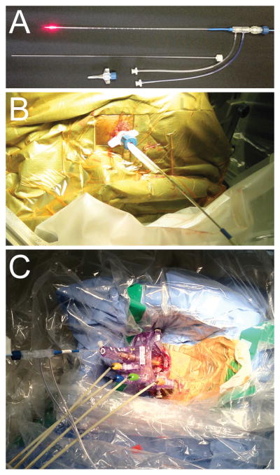
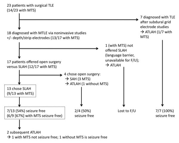
Methods: With patients under general anesthesia and using standard stereotactic methods, 13 adult patients with intractable mesial temporal lobe epilepsy (with and without mesial temporal sclerosis [MTS]) prospectively underwent insertion of a saline-cooled fiberoptic laser applicator in amygdalohippocampal structures from an occipital trajectory. Computer-controlled laser ablation was performed during continuous magnetic resonance thermal imaging followed by confirmatory contrast-enhanced anatomic imaging and volumetric reconstruction. Clinical outcomes were determined from seizure diaries.

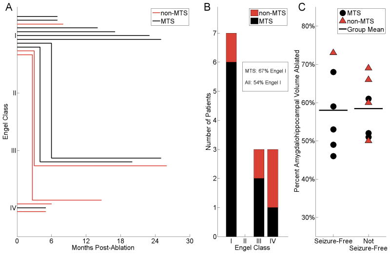
Results: A mean 60% volume of the amygdalohippocampal complex was ablated in 13 patients (9 with MTS) undergoing 15 procedures. Median hospitalization was 1 day. With follow-up ranging from 5 to 26 months (median, 14 months), 77% (10/13) of patients achieved meaningful seizure reduction, of whom 54% (7/13) were free of disabling seizures. Of patients with preoperative MTS, 67% (6/9) achieved seizure freedom. All recurrences were observed before 6 months. Variances in ablation volume and length did not account for individual clinical outcomes. Although no complications of laser therapy itself were observed, 1 significant complication, a visual field defect, resulted from deviated insertion of a stereotactic aligning rod, which was corrected before ablation.

Conclusion: Real-time magnetic resonance-guided stereotactic laser amygdalohippocampotomy is a technically novel, safe, and effective alternative to open surgery. Further evaluation with larger cohorts over time is warranted.


Conflict of interest statement

The terms of this arrangement have been reviewed and approved by Emory University in accordance with its conflict of interest policies.

Figures

Figure 1
Figure 1. Laser thermal therapy system for stereotactic targeting and focused ablation
A, A 15 W 980 nm diode laser (Visualase®) is directed through a saline-cooled laser applicator (top), consisting of a 400 μm core silica optical fiber, capped with a cylindrical diffusing tip (red). The optical fiber is housed within a 1.65 mm diameter polycarbonate saline-cooled cannula. A threaded polycarbonate bone anchor (bottom) is placed over a stiffening stylet (middle) via various stereotactic methods to target deep brain structures. B, Targeting of the laser applicator from an occipital approach with a bone anchor and a CRW stereotactic frame (Integra®). C, Use of an MRI-guided stereotactic trajectory frame (Clearpoint SmartFrame®) with the Visualase® applicator enables implantation and real-time localization of the laser applicator within an MRI suite.
Figure 2
Figure 2. Illustrative treatment cycle
A, Patient 4 was stereotactically implanted from an occipital approach along the long axis of the amygdalohippocampal complex. Using the Visualase® workstation, multiple points (three shown in this plane) were demarcated for intraoperative MRI thermal measurement – red circle (in the ablation zone), white square (anterior mesencephalon), and blue diamond (lateral mesencephalon). B, Measurement of the temperature at the three points during the ablation. Pink hashed area indicates ablation temperatures >90°C, above which thermal spread is considered unpredictable,, and which triggers cycle termination by the workstation. Note that the brainstem temperature does not increase during the procedure. Real-time temperature measurements (left) and estimated ablation areas (right) at 25 seconds (C), 75 seconds (D), and 130 seconds (E). F, After retracting the laser applicator and ablating at a second site, the final estimate of the total ablated area was calculated. Immediate post-operative T1-weighted imaging with contrast highlights the borders of the lesion in the axial (G, red arrow) and coronal (H) planes. I-J, 6 months after the procedure, the amygdalohippocampal complex demonstrates well-circumscribed non-enhancing pseudocystic atrophy.
Figure 3
Figure 3. Volumetric reconstruction of ablation zone with respect to amygdala and hippocampus
A, Axial segmentation of the amygdala (yellow boundary), hippocampus (green), and ablation zone (red) of patient 4. Laser applicator trajectory (blue and white dashed line) is also marked. B, Sagittal segmentation of the same tissue, as well as the applicator track (blue). C, 3-D reconstruction of the amygdala (yellow) and hippocampus (green), as well as the path of the fiber optic (blue) through the tissue. D, 3-D reconstruction from segmented sections of the ablated tissue (red) within the amygdala and hippocampus.
Figure 4
Figure 4. H&E stained pathological specimens resected from mesial temporal lobes of patients following stereotactic laser ablations
A, Section of ablated hippocampus of patient 5, resected during an open surgery 5 months following SLAH procedure, showing complete infarction. Note the absence of viable nuclei and the presence of congested vessels with mildly thickened vascular walls. B, Section of adjacent intact parahippocampal gyrus of patient 5 with normal cellularity and microvasculature. C, Section of en bloc specimen from the parahippocampal region of another patient, resected 3 months following stereotactic laser ablation of the parahippocampal gyrus. Note the cortical layers and the distinct pial boundary (thin black arrows), which contains several blood vessels (block blue arrows) demarcating an intact neocortical gyrus from the ablated parahippocampal gyrus. White square outline marks the area further magnified in D. D, Higher magnification image from C demonstrating perinecrotic microglial infiltrate at the margin of the infarcted tissue (yellow arrowhead). Again note the pial boundary (thin black arrows) containing blood vessels (block blue arrows) between intact and ablated tissues. Subpial gliosis was noted in this and other sections of the specimen, consistent with chronic epilepsy. Scale bars in inferior right corners of each panel.
Figure 5
Figure 5. Patient outcomes with respect to time, preoperative MRI, and postoperative amygdalohippocampal ablation volume
A, Patient outcome in Engel class (I, free of disabling seizures; II, rare disabling seizures; III, worthwhile improvement; IV, no worthwhile improvement) is presented over time following each of 14 ablation procedures in 13 patients with adequate follow-up. Note that all failures to maintain seizure freedom (Engel I) were apparent by <6 months. Thus, seizure freedom beyond 6 months was a predictor of ongoing therapeutic persistence in our cohort. Three subjects with immediate Engel IV outcomes and <6 months presented went on to undergo additional procedures. B, Summary of the number of patients in each Engel outcome class and subdivided by preoperative MRI findings (MTS or non-MTS). Seven of all 13 (54%) patients and 6/9 (67%) of the select MTS patients were Engel I at last follow-up (median 14 months). Only 1/4 (25%) of non-MTS patients maintained Engel I outcome. C, Patient outcomes (seizure-free versus not seizure-free) with respect to proportional volume of the amygdalohippocampal complex that was acutely ablated. Individual subjects are denoted by symbols (black circles, MTS; red triangles, non-MTS); bold horizontal lines indicate group means. The ablation volumes achieved did not appear to predict seizure freedom. MTS, mesial temporal sclerosis, coded in black (lines, bars, and symbols); non-MTS coded in red.
Figure 6
Figure 6. Surgical treatment and outcome of temporal lobe epilepsy patients
Flowchart of 23 patients who underwent temporal lobe surgery for nonlesional epilepsy and MTS over 2.5 years. Seven underwent craniotomies for intracranial subdural grid electrode studies followed by open ATLAH. Apart from one patient unable to provide research consent due to a language barrier, the remaining 18 patients with MTLE were offered the choice of open temporal lobe surgery versus SLAH. 4 of these chose open surgery (3 with MTS), and 13 selected SLAH (9 with MTS). The SLAH cohort represents a ‘real world’ sample of temporal lobe epilepsy patients presenting for surgical consideration.

Comment in

References

    1. Wiebe S, Blume WT, Girvin JP, Eliasziw M. A randomized, controlled trial of surgery for temporal-lobe epilepsy. N Engl J Med. 2001;345:311–318. - PubMed
    1. Spencer S, Huh L. Outcomes of epilepsy surgery in adults and children. Lancet Neurol. 2008;7:525–537. - PubMed
    1. Lutz MT, Clusmann H, Elger CE, Schramm J, Helmstaedter C. Neuropsychological outcome after selective amygdalohippocampectomy with transsylvian versus transcortical approach: a randomized prospective clinical trial of surgery for temporal lobe epilepsy. Epilepsia. 2004;45:809–816. - PubMed
    1. Von Rhein B, et al. Neuropsychological outcome after selective amygdalohippocampectomy: subtemporal versus transsylvian approach. J Neurol Neurosurg Psychiatr. 2012;83:887–893. - PubMed
    1. Crane J, Milner B. Do I know you? Face perception and memory in patients with selective amygdalo-hippocampectomy. Neuropsychologia. 2002;40:530–538. - PubMed

Publication types