Review
. 2014 Jul;75(1):61-71.
doi: 10.1227/NEU.0000000000000344.

Trends in fluorescence image-guided surgery for gliomas

Affiliations
Review

Trends in fluorescence image-guided surgery for gliomas

Jonathan T C Liu et al. Neurosurgery. 2014 Jul.

Abstract

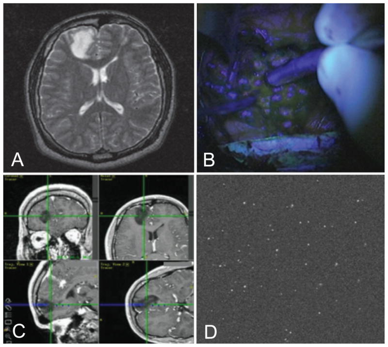
Mounting evidence suggests that a more extensive surgical resection is associated with an improved life expectancy for both low-grade and high-grade glioma patients. However, radiographically complete resections are not often achieved in many cases because of the lack of sensitivity and specificity of current neurosurgical guidance techniques at the margins of diffuse infiltrative gliomas. Intraoperative fluorescence imaging offers the potential to improve the extent of resection and to investigate the possible benefits of resecting beyond the radiographic margins. Here, we provide a review of wide-field and high-resolution fluorescence-imaging strategies that are being developed for neurosurgical guidance, with a focus on emerging imaging technologies and clinically viable contrast agents. The strengths and weaknesses of these approaches will be discussed, as well as issues that are being addressed to translate these technologies into the standard of care.


Conflict of interest statement

DISCLOSURE: The authors report no conflict of interest concerning the surgical methods reviewed in this paper.

Figures

Figure 1
Figure 1
(A) MRI of low-grade glioma. (B) Wide-field FIGS with undetectable fluorescence. (C) Intraoperative neuronavigation showing the location of a confocal microscope within the tumor cavity. (D) Multiple fluorescent cells (> 30) are visualized within the 450 × 450 μm field of view of a miniature confocal microscope.
Figure 2
Figure 2
Example images obtained using advanced FIGS systems. (A–C) Sentinel lymph node (SLN) mapping using the FLARE™ system. (A) White-light image of SLNs. (B) 800 nm NIR fluorescence image following injection of 10 μM ICG:HSA (C) Pseudo-colored (lime green) overlay of the previous two (A,B) images. (D–F) Postmortem imaging of the surgically exposed mouse abdominal area, demonstrating light absorption correction. (D) White-light image. The double arrow indicates the inferior vena cava, while single-line arrows point to the lumbar lymph nodes. (E) Conventional fluorescence image showing poor contrast between lymph nodes and background, following injection of 8 μM Alexa Fluor 750. (F) Normalized/corrected image showing improved fluorescence contrast and quantification of signal from lymph nodes. (G–I) Quantitative fluorescence imaging (qFI) of a human GBM during surgery. Three hours prior to surgery, patient had received 20 mg/kg of ALA. (G) White-light image at the beginning of surgery. (H) Corresponding conventional fluorescence image. (I) Quantitative fluorescence image overlaid with the white-light view.
Figure 3
Figure 3
Representative fluorescent images utilizing passive, metabolic and molecular contrast agents. (A–C) Example images obtained utilizing a passive agent, sodium fluorescein (Yellow 560). (A) Intraoperative white-light image of a metastatic brain tumor (B) Corresponding conventional fluorescence image. (C) Resection of lesion performed with fluorescence image guidance. (D–G) Fluorescence-guided, stepwise resection of a murine RCAS-PDGF glioblastoma, following injection of IRDye 800CW-conjugated RGD peptide (molecular imaging). The tumor (indicated by the black arrowheads) was exposed and a 2-step resection was conducted to remove the glioblastoma tissue (D) White-light image of exposed tumor. (E) Corresponding fluorescence image false-colored and overlaid with a white-light image. (F) Fluorescence image after first resection. (G) Fluorescence image following second resection. (H) Example image using a molecular contrast agent. Raw fluorescence image (false-colored) of a tumor margin stained with a VEGFR-1 probe (785-nm fluorescence excitation). (I) Example image of a metabolic contrast agent. Topical application of the quenched activity-based probe (qABP) GB119 reveals residual tumor in the right side of a mouse brain. The normal tissue on the left side of the brain does not generate strong fluorescence when GB119 is topically applied.(J–M) Dual-reporter imaging of a human-neuronal glioblastoma (U251) tumor in a mouse at 1 hr post injection. (J) White-light image. The white arrows signal the location of the tumor. (K) Corresponding untargeted fluorescence uptake (IRdye 700DX), (L) Targeted fluorescence uptake (EGF-IRdye 800CW), and (M) dual-reporter image of the A431 tumor line. By utilizing a secondary, untargeted imaging reporter, this technique compensates for nonspecific uptake of the targeted fluorophore, hence achieving a more accurate image of molecular expression.
Figure 4
Figure 4
(A) A handheld confocal microscope (Zeiss Optiscan®) used during neurosurgery. (B) Zeiss Optiscan® image revealing localized subcellular 5-ALA-induced tumor fluorescence in a low-grade glioma. (C) Intraoperative confocal image of tumor microvasculature following injection of sodium fluorescein. (D) Miniature dual-axis confocal microscope prototype. (E) Image of GFP-expressing tumor cells in a spontaneous mouse model of medulloblastoma. (F) Fluorescence image of mouse brain vasculature (depth 55 μm) following retro-orbital injection of fluorescein-dextran.

References

    1. Smith JS, Chang EF, Lamborn KR, et al. Role of extent of resection in the long-term outcome of low-grade hemispheric gliomas. J Clin Oncol. 2008;26:1338–1345. - PubMed
    1. Hardesty DA, Sanai N. The value of glioma extent of resection in the modern neurosurgical era. Front Neurol. 2012;3:140. - PMC - PubMed
    1. Duffau H, Mandonnet E. The “onco-functional balance” in surgery for diffuse low-grade glioma: integrating the extent of resection with quality of life. Acta Neurochir (Wien ) 2013;155:951–957. - PubMed
    1. Duffau H. A new concept of diffuse (low-grade) glioma surgery. Adv Tech Stand Neurosurg. 2012;38:3–27. - PubMed
    1. Liao H, Noguchi M, Maruyama T, et al. An integrated diagnosis and therapeutic system using intra-operative 5-aminolevulinic-acid-induced fluorescence guided robotic laser ablation for precision neurosurgery. Med Image Anal. 2012;16:754–766. - PubMed

Publication types