. 2014 Mar 11;9(3):e91574.
doi: 10.1371/journal.pone.0091574. eCollection 2014.

Pro-inflammatory chemokine CCL2 (MCP-1) promotes healing in diabetic wounds by restoring the macrophage response

Affiliations

Pro-inflammatory chemokine CCL2 (MCP-1) promotes healing in diabetic wounds by restoring the macrophage response

Stephen Wood et al. PLoS One. .

Abstract

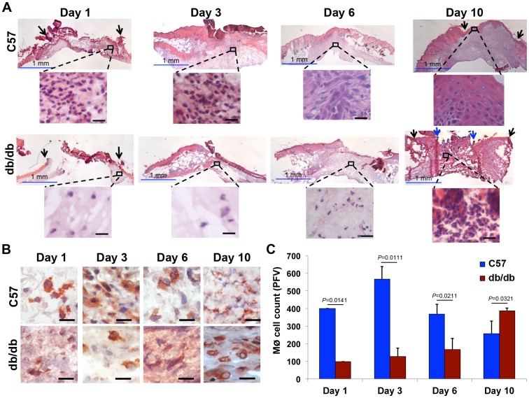
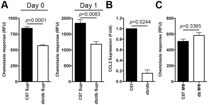
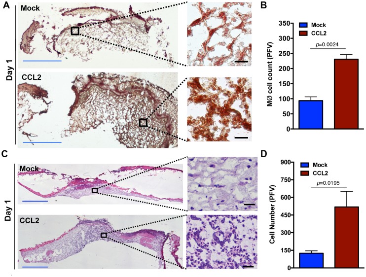
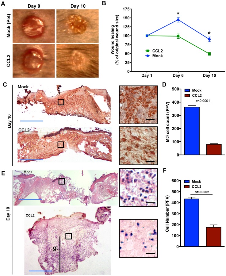
Prior studies suggest that the impaired healing seen in diabetic wounds derives from a state of persistent hyper-inflammation characterized by harmful increases in inflammatory leukocytes including macrophages. However, such studies have focused on wounds at later time points (day 10 or older), and very little attention has been given to the dynamics of macrophage responses in diabetic wounds early after injury. Given the importance of macrophages for the process of healing, we studied the dynamics of macrophage response during early and late phases of healing in diabetic wounds. Here, we report that early after injury, the diabetic wound exhibits a significant delay in macrophage infiltration. The delay in the macrophage response in diabetic wounds results from reduced Chemokine (C-C motif) ligand 2 (CCL2) expression. Importantly, one-time treatment with chemoattractant CCL2 significantly stimulated healing in diabetic wounds by restoring the macrophage response. Our data demonstrate that, rather than a hyper-inflammatory state; the early diabetic wound exhibits a paradoxical and damaging decrease in essential macrophage response. Our studies suggest that the restoration of the proper kinetics of macrophage response may be able to jumpstart subsequent healing stages. CCL2 chemokine-based therapy may be an attractive strategy to promote healing in diabetic wounds.


Conflict of interest statement

Competing Interests: The authors have declared that no competing interests exist.

Figures

Figure 1
Figure 1. Macrophage response is delayed in diabetic skin tissue.
Skin tissues from normal (C57) and diabetic (db/db) wounds were harvested at indicated time-points post wounding (day 0), fixed and stained with hematoxylin and eosin (A) or with CD68 antibody which primarily stains macrophages (B). The corresponding tabulated data for macrophage cell counts are shown as the mean ± SEM in (C) (N = 3 mice for H&E staining in A; n = 18 for B-C. 3 mice per group, 6 random fields per mouse from underneath the wound extending into the provisional matrix in the dermis region, all p-values were significant ranging from 0.0111 to 0.0321). Black arrows point to the original sites of incision. Blue arrows point to leading edges of diabetic wounds on day 10. For clarity and to enhance magnification, representative cropped regions from underneath the wounds extending in the dermis are shown. Combined, the data indicate that inflammatory response is delayed in diabetic tissues. Blue scale bar  =  1 mm, black scale bar  =  25 μm.
Figure 2
Figure 2. Chemokine insufficiency leads to impaired macrophage response in diabetic wounds early after injury.
(A) Skin tissues from the normal and diabetic wound edges (1 mm from the rim) were harvested on day 0 (right after wounding) and on day 1 post wounding. These tissue explants were cultured and allowed to secrete chemokines into culture media. The supernatants from these cultures were then evaluated for their chemokine levels by assessing their ability to chemoattract normal macrophages from C57. The results are shown as the mean ± SEM (n = 18, 6 mice per group, each done in duplicates), indicating that diabetic supernatants contain significantly reduced chemoattractants for macrophages (MΦs). (B) CCL2 chemokine expression levels in day 1 C57 and db/db wounds were determined by RT-PCR and the results are shown as the mean ± SEM, indicating significant reduction in day 1 diabetic wounds (n = 3 mice per group). (C) Diabetic and normal MΦs were evaluated for their ability to chemotax toward CCL2, a known chemoattractant for MΦs. The results are shown as the mean ± SEM (n = 6, p = 0.3365), indicating that there is no significant differences in chemotactic responses between normal and diabetic MΦs toward CCL2.
Figure 3
Figure 3. CCL2 treatment enhances monocytes infiltration in diabetic wound.
Diabetic wounds were treated with PBS (Mock) or PBS + CCL2 (CCL2). Treated wound tissues were harvested on day 1 after treatment and stained with a macrophage/monocyte-specific antibody (CD68) in (A) or H&E in (C). The corresponding tabulated data for (A) and (C) are shown in (B) and (D) as mean ± SEM (N = 18, 3 mice, 6 random fields from the dermal region, p = 0.0024 for B and p = 0.0195 for D). For clarity and to enhance magnification, representative cropped regions from underneath the wounds extending in the dermis are shown. Blue scale bar  =  1 mm, black scale bar  =  25 μm.
Figure 4
Figure 4. CCL2 treatment stimulates healing in diabetic wound.
(A) Wound healing in the mock and CCL2-treated diabetic wounds was monitored by digital photography and wound areas were determined by tracing. Representative images from days 0 and 10 are shown in (A) and the tabulated results are shown as mean ± SEM in (B) (* signifies significance with p<0.0001, n = 4). Skin tissues from mock- or CCL2-treated diabetic wounds were harvested on day 10 after treatment, stained with monocyte/macrophage antibody (CD68) (C) or H&E (E). The corresponding tabulated data for (C) and (E) are shown in (D) and (F) as mean ± SEM (N = 18, 3 mice, 6 random fields from the dermal region, p<0.0001 for D and p = 0.0002 for F). As indicated, day 10 CCL2-treated diabetic wounds exhibit enhanced healing, complete re-epithelization, epidermal thickening, increased granulation tissue (as indicated by gt), and reduced inflammation compared to mock-treated diabetic wounds which are partially re-epithelized and are highly inflamed. For clarity and to enhance magnification, representative cropped regions from underneath the wounds extending in the dermis are shown. Blue scale bar  =  1 mm, black scale bar  =  25 μm.

References

    1. Singer AJ, Clark RA (1999) Cutaneous wound healing. N Engl J Med 341: 738–746. - PubMed
    1. Bjarnsholt T, Kirketerp-Moller K, Jensen PO, Madsen KG, Phipps R, et al. (2008) Why chronic wounds will not heal: a novel hypothesis. Wound Repair Regen 16: 2–10. - PubMed
    1. Donath MY, Shoelson SE (2011) Type 2 diabetes as an inflammatory disease. Nat Rev Immunol 11: 98–107. - PubMed
    1. Shaw TJ, Martin P (2009) Wound repair at a glance. J Cell Sci 122: 3209–3213. - PMC - PubMed
    1. Adzick NS, Harrison MR, Glick PL, Beckstead JH, Villa RL, et al. (1985) Comparison of fetal, newborn, and adult wound healing by histologic, enzyme-histochemical, and hydroxyproline determinations. J Pediatr Surg 20: 315–319. - PubMed

Publication types