Review
. 2014:807:75-96.
doi: 10.1007/978-81-322-1777-0_6.

Neuroinvasive and neurotropic human respiratory coronaviruses: potential neurovirulent agents in humans

Affiliations
Review

Neuroinvasive and neurotropic human respiratory coronaviruses: potential neurovirulent agents in humans

Marc Desforges et al. Adv Exp Med Biol. 2014.

Abstract

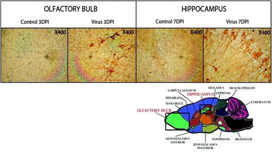
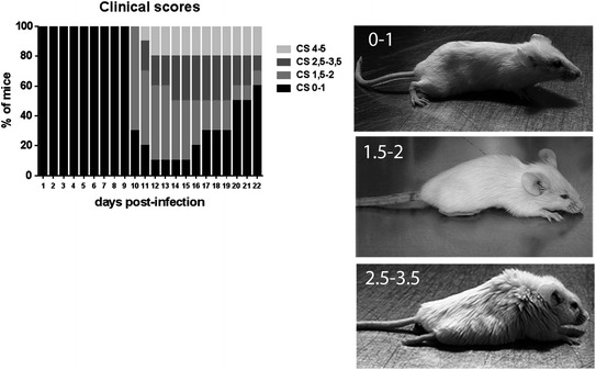
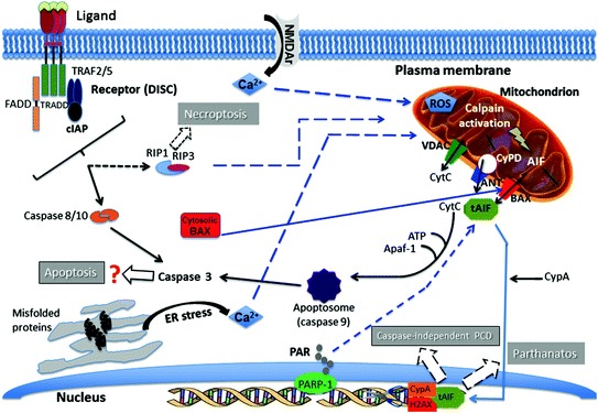
In humans, viral infections of the respiratory tract are a leading cause of morbidity and mortality worldwide. Several recognized respiratory viral agents have a neuroinvasive capacity since they can spread from the respiratory tract to the central nervous system (CNS). Once there, infection of CNS cells (neurotropism) could lead to human health problems, such as encephalitis and long-term neurological diseases. Among the various respiratory viruses, coronaviruses are important pathogens of humans and animals. Human Coronaviruses (HCoV) usually infect the upper respiratory tract, where they are mainly associated with common colds. However, in more vulnerable populations, such as newborns, infants, the elderly, and immune-compromised individuals, they can also affect the lower respiratory tract, leading to pneumonia, exacerbations of asthma, respiratory distress syndrome, or even severe acute respiratory syndrome (SARS). The respiratory involvement of HCoV has been clearly established since the 1960s. In addition, for almost three decades now, the scientific literature has also demonstrated that HCoV are neuroinvasive and neurotropic and could induce an overactivation of the immune system, in part by participating in the activation of autoreactive immune cells that could be associated with autoimmunity in susceptible individuals. Furthermore, it was shown that in the murine CNS, neurons are the main target of infection, which causes these essential cells to undergo degeneration and eventually die by some form of programmed cell death after virus infection. Moreover, it appears that the viral surface glycoprotein (S) represents an important factor in the neurodegenerative process. Given all these properties, it has been suggested that these recognized human respiratory pathogens could be associated with the triggering or the exacerbation of neurological diseases for which the etiology remains unknown or poorly understood.


Figures

Fig. 1
Fig. 1
Illustration of the transneuronal route used by HCoV-OC43 for neuroinvasion and dissemination into the central nervous system. The left panel shows the olfactory bulb area, either mock-infected (control) or HCoV-OC43-infected (virus) at 3 days postinfection (DPI). The right panel shows the hippocampus, either mock-infected (control) or HCoV-OC43-infected (virus) at 7 days postinfection (DPI). In both regions of the brain, neurons are the target of infection. Magnification is 400X
Fig. 2
Fig. 2
Evaluation of the clinical scores (CS) related to motor dysfunctions (paralysis) of HCoV-OC43 infected mice. The left panel shows the percentage of mice observed at each of the different degree of motor dysfunctions over the course of infection, from 0 to 21 days postinfection. The CS were established according to a scale based on the recognized experimental allergic encephalitis (EAE) model (01 normal mouse with no clinical signs; 1.52 partial hind-limb paralysis, with a walk close to ground level; 2.53.5 complete hind-limb paralysis, and 45 moribund state or death. The right panels are a representation of mice at each stage of paralysis
Fig. 3
Fig. 3
Pathways of neuronal degeneration and cell death induced by HCoV–OC43 infection. Several cellular factors that regulate various mechanisms are activated in response to infection, which leads to programmed cell death (PCD). (1) Hallmarks of apoptosis, including relocalization of the activated pro-apoptotic protein BAX (Bcl-2 associated protein X) from the cytosol to the mitochondrial membrane, cytochrome C release from mitochondria toward the cytosol, DNA fragmentation, and activation of caspases -3 and -9, are observed during infection of human neurons. However, using a pan-caspase inhibitor (Z-VAD-fmk), cell death is not abrogated after infection, suggesting a caspase-independent type of apoptosis. (2) Relocalization of the mitochondrial protein AIF (apoptosis-inducing factor) toward the nucleus (tAIF) is observed after infection and participates in DNA fragmentation in conjunction with CypA (cyclophilin A) and histone H2AX. The AIF is known to be activated during caspase-independent apoptosis. However, AIF is also involved in Parthanatos, another form of PCD potentially associated with neurodegeneration. As they are synthesized by the poly(ADP-ribose) polymerase (PARP) during a neuronal stress, including during HCoV–OC43 infection, polymers of ADP-ribose (PAR) may relocalize toward mitochondria and participate in the activation and relocalization of AIF toward the cytosol before it reaches the nucleus. Cyclophilin D (CypD) inhibition decreases AIF release from mitochondria and abrogates cell death induced by infection. (3) AIF release from mitochondria may be induced through its truncation (tAIF) by activated calpain, which is usually activated by a rise in the mitochondrial calcium concentration. (4) This increase in calcium concentration may be linked with either an important entry from the extracellular milieu (for instance during excitotoxicity) or with a release of calcium from the endoplasmic reticulum (ER) following induction of ER stress. Both situations are probably taking place after infection of neurons by HCoV-OC43. The increase in calcium concentration in mitochondria may also induce production of reactive oxygen species (ROS) that can be harmful for mitochondria and hence neurons. (5) The presence during infection of an inhibitor (Nec-1) of the receptor interacting protein kinase-1 (RIP-1), significantly increases cell survival and partially abrogates viral replication, suggesting that necroptosis, a third form of PCD which involves RIP-1 and RIP-3 downstream of the tumor necrosing factor (TNF) receptor family (in the form of the death-inducing signaling complex (DISC)), may play a role in HCoV-OC43-induced neuronal death. Solid arrows indicate experimental data and dashed arrows represent possible pathways based on the current literature (see text for details)

References

    1. Vareille M, Kieninger E, Edwards MR, Regamey N. The airway epithelium: soldier in the fight against respiratory viruses. Clin Microbiol Rev. 2011;24(1):210–229. - PMC - PubMed
    1. Jartti T, Jartti L, Ruuskanen O, Soderlund-Venermo M. New respiratory viral infections. Curr Opin Pulm Med. 2012;18(3):271–278. - PubMed
    1. Ison MG, Hayden FG. Viral infections in immunocompromised patients: what’s new with respiratory viruses? Curr Opin Infect Dis. 2002;15(4):355–367. - PubMed
    1. Cesario TC. Viruses associated with pneumonia in adults. Clin Infect Dis. 2012;55(1):107–113. - PMC - PubMed
    1. Sloots TP, Whiley DM, Lambert SB, Nissen MD. Emerging respiratory agents: new viruses for old diseases? J Clin Virol. 2008;42(3):233–243. - PMC - PubMed

LinkOut - more resources