

Computerized image analysis for identifying triple-negative breast cancers and differentiating them from other molecular subtypes of breast cancer on dynamic contrast-enhanced MR images: a feasibility study
- PMID: 24620909
- PMCID: PMC4263619
- DOI: 10.1148/radiol.14121031
Computerized image analysis for identifying triple-negative breast cancers and differentiating them from other molecular subtypes of breast cancer on dynamic contrast-enhanced MR images: a feasibility study
Abstract
Purpose: To determine the feasibility of using a computer-aided diagnosis (CAD) system to differentiate among triple-negative breast cancer, estrogen receptor (ER)-positive cancer, human epidermal growth factor receptor type 2 (HER2)-positive cancer, and benign fibroadenoma lesions on dynamic contrast material-enhanced (DCE) magnetic resonance (MR) images.
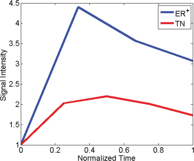
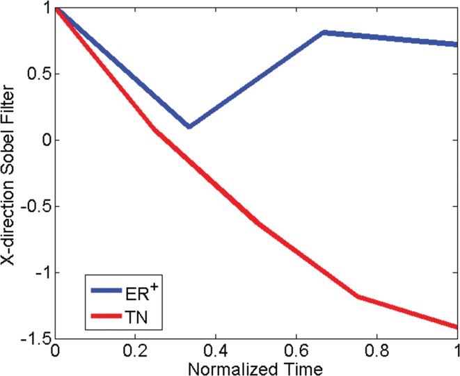
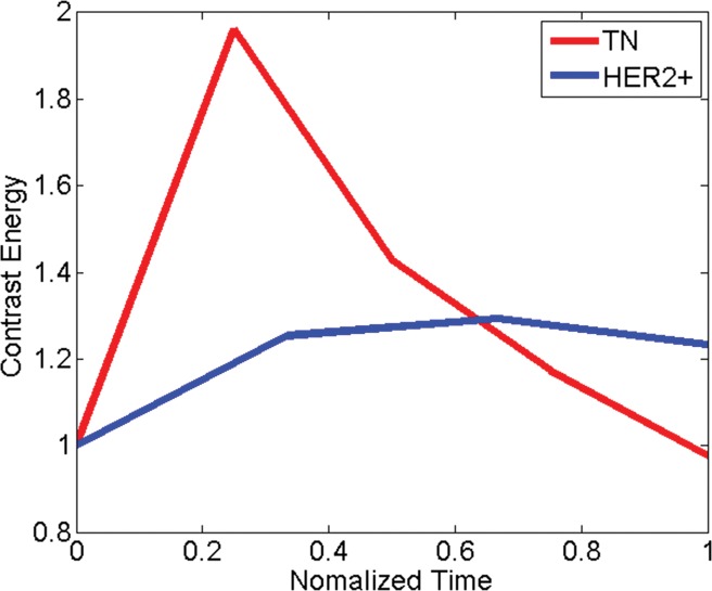
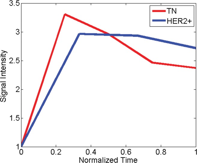
Materials and methods: This is a retrospective study of prospectively acquired breast MR imaging data collected from an institutional review board-approved, HIPAA-compliant study between 2002 and 2007. Written informed consent was obtained from all patients. The authors collected DCE MR images from 65 women with 76 breast lesions who had been recruited into a larger study of breast MR imaging. The women had triple-negative (n = 21), ER-positive (n = 25), HER2-positive (n = 18), or fibroadenoma (n = 12) lesions. All lesions were classified as Breast Imaging Reporting and Data System category 4 or higher on the basis of previous imaging. Images were subject to quantitative feature extraction, feed-forward feature selection by means of linear discriminant analysis, and lesion classification by using a support vector machine classifier. The area under the receiver operating characteristic curve (Az) was calculated for each of five lesion classification tasks involving triple-negative breast cancers.
Results: For each pair-wise lesion type comparison, linear discriminant analysis helped identify the most discriminatory features, which in conjunction with a support vector machine classifier yielded an Az of 0.73 (95% confidence interval [CI]: 0.59, 0.87) for triple-negative cancer versus all non-triple-negative lesions, 0.74 (95% CI: 0.60, 0.88) for triple-negative cancer versus ER- and HER2-positive cancer, 0.77 (95% CI: 0.63, 0.91) for triple-negative versus ER-positive cancer, 0.74 (95% CI: 0.58, 0.89) for triple-negative versus HER2-positive cancer, and 0.97 (95% CI: 0.91, 1.00) for triple-negative cancer versus fibroadenoma.
Conclusion: Triple-negative cancers possess certain characteristic features on DCE MR images that can be captured and quantified with CAD, enabling good discrimination of triple-negative cancers from non-triple-negative cancers, as well as between triple-negative cancers and benign fibroadenomas. Such CAD algorithms may provide added diagnostic benefit in identifying the highly aggressive triple-negative cancer phenotype with DCE MR imaging in high-risk women.
© RSNA, 2014.
Figures
Similar articles
-
Cancerous breast lesions on dynamic contrast-enhanced MR images: computerized characterization for image-based prognostic markers.Radiology. 2010 Mar;254(3):680-90. doi: 10.1148/radiol.09090838. Epub 2010 Feb 1. Radiology. 2010. PMID: 20123903 Free PMC article.
-
Improving the Accuracy of Computer-aided Diagnosis for Breast MR Imaging by Differentiating between Mass and Nonmass Lesions.Radiology. 2016 Mar;278(3):679-88. doi: 10.1148/radiol.2015150241. Epub 2015 Sep 18. Radiology. 2016. PMID: 26383229
-
Identifying Triple-Negative Breast Cancer Using Background Parenchymal Enhancement Heterogeneity on Dynamic Contrast-Enhanced MRI: A Pilot Radiomics Study.PLoS One. 2015 Nov 24;10(11):e0143308. doi: 10.1371/journal.pone.0143308. eCollection 2015. PLoS One. 2015. PMID: 26600392 Free PMC article.
-
Triple-Negative Breast Cancer: Radiologic-Pathologic Correlation.J Breast Imaging. 2025 May 17;7(3):331-344. doi: 10.1093/jbi/wbae085. J Breast Imaging. 2025. PMID: 39801352 Review.
-
Breast Cancer Subtypes and Quantitative Magnetic Resonance Imaging: A Systemic Review.Life (Basel). 2022 Mar 28;12(4):490. doi: 10.3390/life12040490. Life (Basel). 2022. PMID: 35454981 Free PMC article. Review.
Cited by
-
Nodal-based radiomics analysis for identifying cervical lymph node metastasis at levels I and II in patients with oral squamous cell carcinoma using contrast-enhanced computed tomography.Eur Radiol. 2021 Oct;31(10):7440-7449. doi: 10.1007/s00330-021-07758-4. Epub 2021 Mar 31. Eur Radiol. 2021. PMID: 33787970 Clinical Trial.
-
Multiparametric Analysis of Longitudinal Quantitative MRI data to Identify Distinct Tumor Habitats in Preclinical Models of Breast Cancer.Cancers (Basel). 2020 Jun 24;12(6):1682. doi: 10.3390/cancers12061682. Cancers (Basel). 2020. PMID: 32599906 Free PMC article.
-
Unsupervised Clustering of Quantitative Image Phenotypes Reveals Breast Cancer Subtypes with Distinct Prognoses and Molecular Pathways.Clin Cancer Res. 2017 Jul 1;23(13):3334-3342. doi: 10.1158/1078-0432.CCR-16-2415. Epub 2017 Jan 10. Clin Cancer Res. 2017. PMID: 28073839 Free PMC article.
-
A Radio-genomics Approach for Identifying High Risk Estrogen Receptor-positive Breast Cancers on DCE-MRI: Preliminary Results in Predicting OncotypeDX Risk Scores.Sci Rep. 2016 Feb 18;6:21394. doi: 10.1038/srep21394. Sci Rep. 2016. PMID: 26887643 Free PMC article. Clinical Trial.
-
DCE-MRI radiomics of primary breast lesions combined with ipsilateral axillary lymph nodes for predicting efficacy of NAT.BMC Cancer. 2025 Apr 1;25(1):589. doi: 10.1186/s12885-025-14004-3. BMC Cancer. 2025. PMID: 40170181 Free PMC article.
References
-
- Reis-Filho JS, Tutt ANJ. Triple negative tumours: a critical review. Histopathology 2008;52(1):108–118. - PubMed
-
- Stockmans G, Deraedt K, Wildiers H, Moerman P, Paridaens R. Triple-negative breast cancer. Curr Opin Oncol 2008;20(6):614–620. - PubMed
-
- Haffty BG, Yang Q, Reiss M, et al. . Locoregional relapse and distant metastasis in conservatively managed triple negative early-stage breast cancer. J Clin Oncol 2006;24(36):5652–5657. - PubMed
-
- Schrading S, Kuhl CK. Mammographic, US, and MR imaging phenotypes of familial breast cancer. Radiology 2008;246(1):58–70. - PubMed
-
- Tchou J, Wang LP, Sargen M, Sonnad S, Tomaszewski J, Schnall M. Do triple-negative breast cancers have a distinct imaging phenotype? Presented at the 29th Annual San Antonio Breast Cancer Symposium, San Antonio, Tex, December 14–17, 2006.
MeSH terms
Substances
Grants and funding
LinkOut - more resources
Full Text Sources
Other Literature Sources
Medical
Research Materials
Miscellaneous
