

Computerized image analysis for identifying triple-negative breast cancers and differentiating them from other molecular subtypes of breast cancer on dynamic contrast-enhanced MR images: a feasibility study
- PMID: 24620909
- PMCID: PMC4263619
- DOI: 10.1148/radiol.14121031
Computerized image analysis for identifying triple-negative breast cancers and differentiating them from other molecular subtypes of breast cancer on dynamic contrast-enhanced MR images: a feasibility study
Abstract
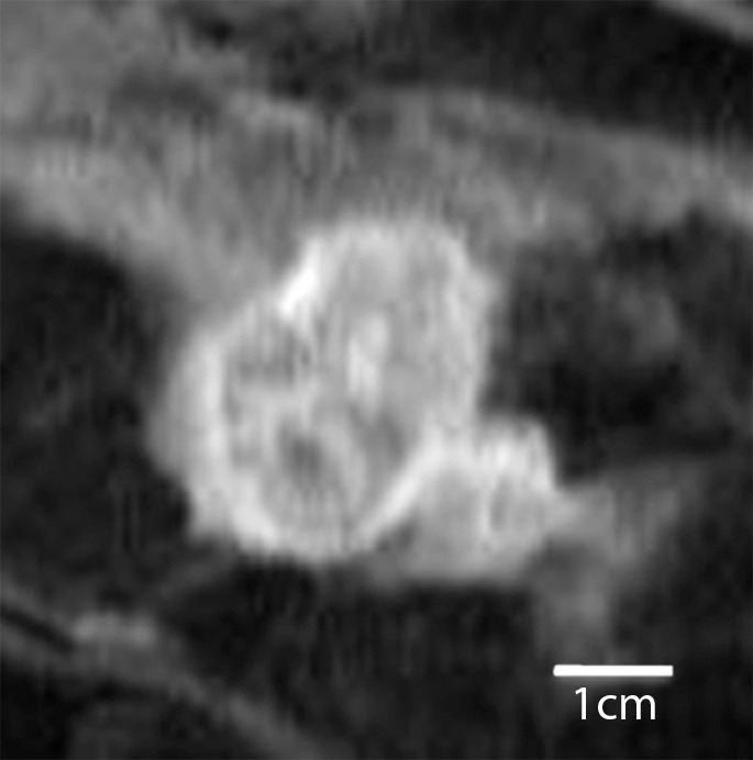
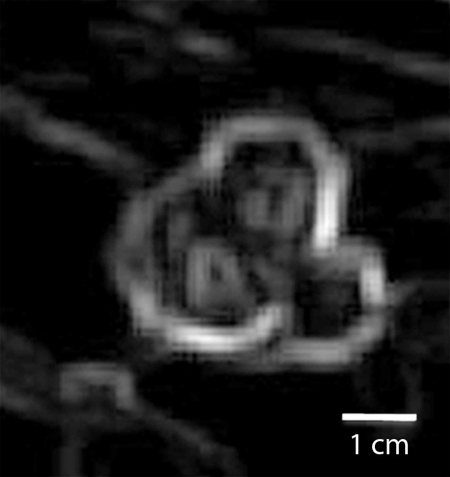
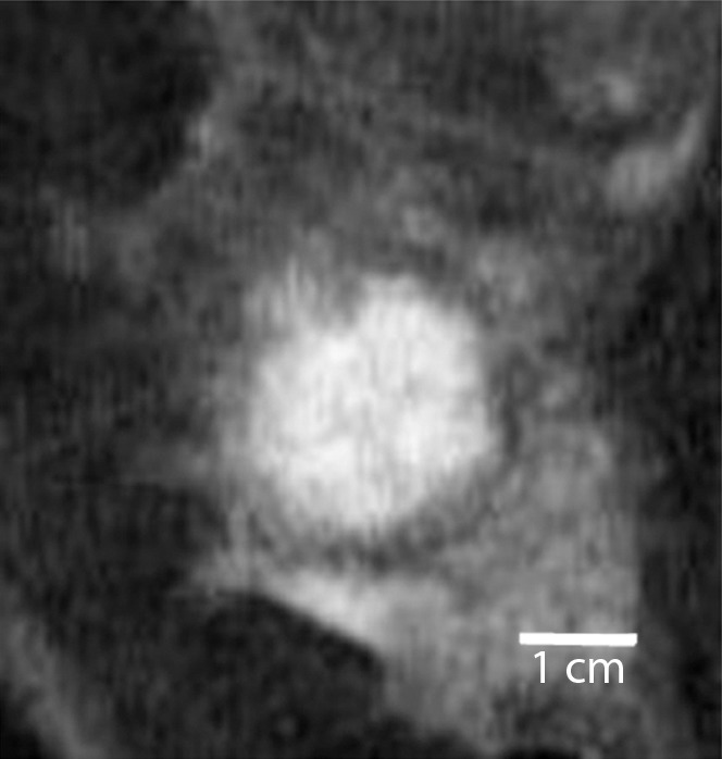
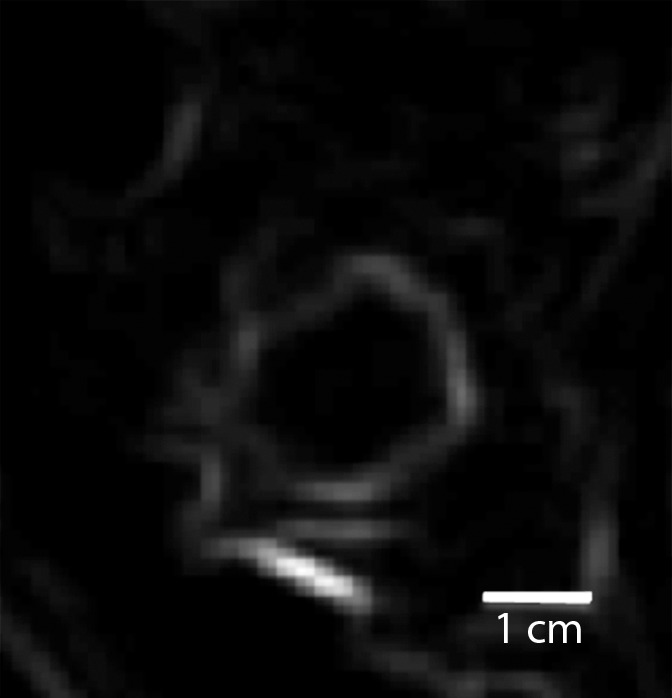
Purpose: To determine the feasibility of using a computer-aided diagnosis (CAD) system to differentiate among triple-negative breast cancer, estrogen receptor (ER)-positive cancer, human epidermal growth factor receptor type 2 (HER2)-positive cancer, and benign fibroadenoma lesions on dynamic contrast material-enhanced (DCE) magnetic resonance (MR) images.
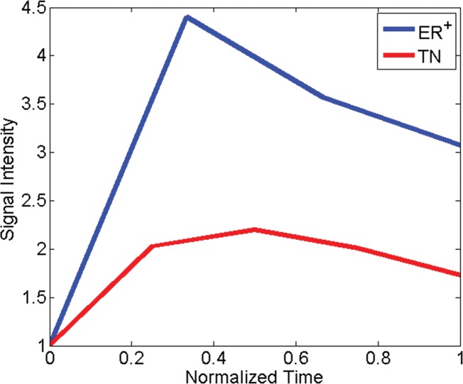
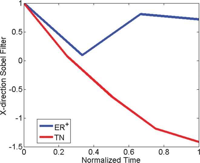
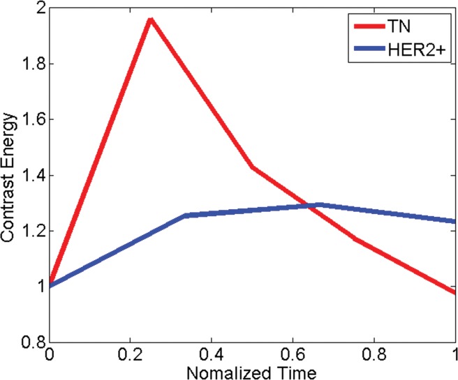
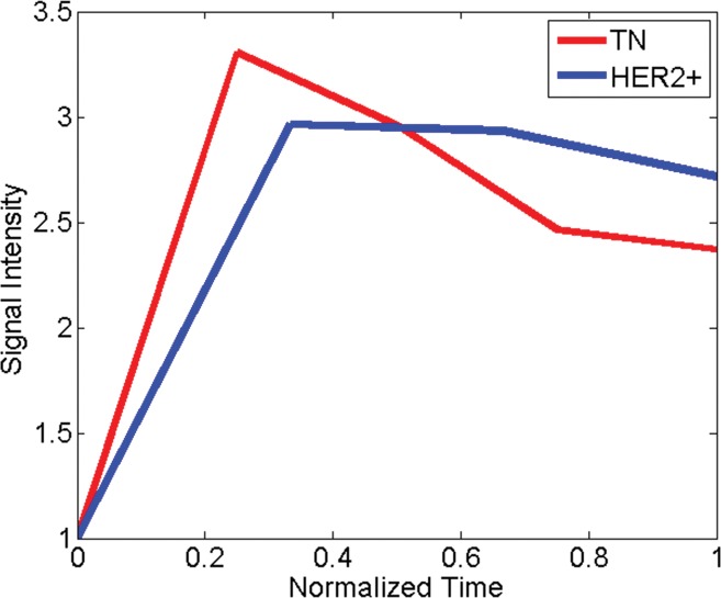
Materials and methods: This is a retrospective study of prospectively acquired breast MR imaging data collected from an institutional review board-approved, HIPAA-compliant study between 2002 and 2007. Written informed consent was obtained from all patients. The authors collected DCE MR images from 65 women with 76 breast lesions who had been recruited into a larger study of breast MR imaging. The women had triple-negative (n = 21), ER-positive (n = 25), HER2-positive (n = 18), or fibroadenoma (n = 12) lesions. All lesions were classified as Breast Imaging Reporting and Data System category 4 or higher on the basis of previous imaging. Images were subject to quantitative feature extraction, feed-forward feature selection by means of linear discriminant analysis, and lesion classification by using a support vector machine classifier. The area under the receiver operating characteristic curve (Az) was calculated for each of five lesion classification tasks involving triple-negative breast cancers.
Results: For each pair-wise lesion type comparison, linear discriminant analysis helped identify the most discriminatory features, which in conjunction with a support vector machine classifier yielded an Az of 0.73 (95% confidence interval [CI]: 0.59, 0.87) for triple-negative cancer versus all non-triple-negative lesions, 0.74 (95% CI: 0.60, 0.88) for triple-negative cancer versus ER- and HER2-positive cancer, 0.77 (95% CI: 0.63, 0.91) for triple-negative versus ER-positive cancer, 0.74 (95% CI: 0.58, 0.89) for triple-negative versus HER2-positive cancer, and 0.97 (95% CI: 0.91, 1.00) for triple-negative cancer versus fibroadenoma.
Conclusion: Triple-negative cancers possess certain characteristic features on DCE MR images that can be captured and quantified with CAD, enabling good discrimination of triple-negative cancers from non-triple-negative cancers, as well as between triple-negative cancers and benign fibroadenomas. Such CAD algorithms may provide added diagnostic benefit in identifying the highly aggressive triple-negative cancer phenotype with DCE MR imaging in high-risk women.
© RSNA, 2014.
Figures
References
-
- Reis-Filho JS, Tutt ANJ. Triple negative tumours: a critical review. Histopathology 2008;52(1):108–118. - PubMed
-
- Stockmans G, Deraedt K, Wildiers H, Moerman P, Paridaens R. Triple-negative breast cancer. Curr Opin Oncol 2008;20(6):614–620. - PubMed
-
- Haffty BG, Yang Q, Reiss M, et al. Locoregional relapse and distant metastasis in conservatively managed triple negative early-stage breast cancer. J Clin Oncol 2006;24(36):5652–5657. - PubMed
-
- Schrading S, Kuhl CK. Mammographic, US, and MR imaging phenotypes of familial breast cancer. Radiology 2008;246(1):58–70. - PubMed
-
- Tchou J, Wang LP, Sargen M, Sonnad S, Tomaszewski J, Schnall M. Do triple-negative breast cancers have a distinct imaging phenotype? Presented at the 29th Annual San Antonio Breast Cancer Symposium, San Antonio, Tex, December 14–17, 2006.
MeSH terms
Substances
Grants and funding
LinkOut - more resources
Full Text Sources
Other Literature Sources
Medical
Research Materials
Miscellaneous
