Review
. 2014;8(2):125-35.
doi: 10.4161/cam.28243.

Dynamics between actin and the VE-cadherin/catenin complex: novel aspects of the ARP2/3 complex in regulation of endothelial junctions

Affiliations
Review

Dynamics between actin and the VE-cadherin/catenin complex: novel aspects of the ARP2/3 complex in regulation of endothelial junctions

Abdallah Abu Taha et al. Cell Adh Migr. 2014.

Abstract

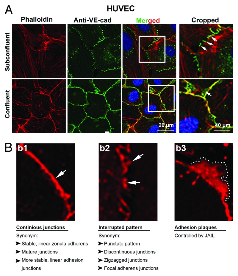
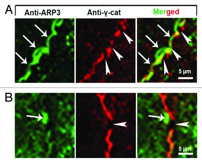
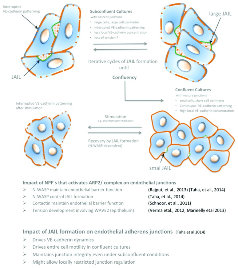
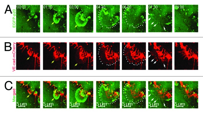
Endothelial adherens junctions are critical for physiological and pathological processes such as differentiation, maintenance of entire monolayer integrity, and the remodeling. The endothelial-specific VE-cadherin/catenin complex provides the backbone of adherens junctions and acts in close interaction with actin filaments and actin/myosin-mediated contractility to fulfill the junction demands. The functional connection between the cadherin/catenin complex and actin filaments might be either directly through ?-catenins, or indirectly e.g., via linker proteins such as vinculin, p120ctn, ?-actinin, or EPLIN. However, both junction integrity and dynamic remodeling have to be contemporarily coordinated. The actin-related protein complex ARP2/3 and its activating molecules, such as N-WASP and WAVE, have been shown to regulate the lammellipodia-mediated formation of cell junctions in both epithelium and endothelium. Recent reports now demonstrate a novel aspect of the ARP2/3 complex and the nucleating-promoting factors in the maintenance of endothelial barrier function and junction remodeling of established endothelial cell junctions. Those mechanisms open novel possibilities; not only in fulfilling physiological demands but obtained information may be of critical importance in pathologies such as wound healing, angiogenesis, inflammation, and cell diapedesis.

Keywords: ARP2/3 complex; VE-cadherin; actin; cortical actin; endothelium; stress fibers.


Figures

None
Figure 1. Distribution of VE-cadherin and actin at cell junctions of subconfluent and confluent endothelial cells in culture. Subconfluent and confluent human umbilical vein endothelial cells (HUVEC) cultures were labeled with (a) anti-VE-cadherin (green) and phalloidin-TRITC for filamentous actin (red) or (b1–3) VE-cadherin alone (red). (a) VE-cadherin appears interrupted in the subconfluent cultures (arrows) with large cells and extended perimeter, while cells of confluent cultures are small and polygonal and preferentially exhibits a continuous VE cadherin distribution (arrow). Boxes in the merged images indicated the cropped area as indicated. Actin filaments are incomplete co-localized with the VE-cadherin/catenin complex in both subconfluent and confluent cultures. (b1–3) Higher magnification of HUVEC cultures labeled with anti-VE-cadherin (red). (b1) VE-cadherin appears in confluent cultures in a continuous VE cadherin patterning (arrow) while (b2) in subconfluent cultures, VE-cadherin preferentially appears as an interrupted patterning by which VE-cadherin cluster display different sizes (arrows). (b3) A VE-cadherin adhesion plaques, which is a result of JAIL formation is indicated (dotted line).
None
Figure 4. Scheme illustrating the interdependency between JAIL-activity and VE-cadherin dynamics in relation to cell density. For details, please compare text.
None
Figure 2. High magnification of cell junctions in (A) subconfluent and (B) confluent HUVEC cultures labeled with antibodies as indicated. (A) JAIL (arrows) preferentially appear at spaces close to and between VE-cadherin/catenin clusters (arrowheads). (B) Even in confluent cultures, small interruptions of the continuous VE-cadherin/catenin line appear spatiotemporally restricted followed by formation of small ARP2/3 complex controlled JAIL. Taken from Taha et al., 2014, MBoC.
None
Figure 3. Time-lapse series of ARP2/3 complex-mediated JAIL formation and VE-cadherin dynamics in subconfluent endothelial cell cultures expressing both the fusion protein EGFP-p20 (green) and VE-cadherin-mCherry (red) at high magnification. (A) JAIL developed (yellow arrows) to its maximal extension within 4–5 min (green, dotted lines). JAIL developed close to and between interruptions of VE-cadherin-m-Cherry clusters and caused new VE-cadherin adhesion plaques (B, yellow arrows and dotted lines). (B) VE-cadherin-mCherry plaques (dotted lines) increasingly cluster (white arrows) during JAIL retraction and assemble at cell junctions. This mechanisms change the VE-cadherin pattern, and thus, contributes to VE-cadherin dynamics. Taken from Taha et al., 2014, MBoC.

References

    1. Dejana E, Vestweber D. The Role VE-Cadherin in Vascular Morphogenesis and Permeability Control. In: VanRoy F, ed. Molecular Biology of Cadherins, 2013:119-44. - PubMed
    1. Nelson WJ, Dickinson DJ, Weis WI. Roles of cadherins and catenins in cell-cell adhesion and epithelial cell polarity. Prog Mol Biol Transl Sci. 2013;116:3–23. doi: 10.1016/B978-0-12-394311-8.00001-7. - DOI - PubMed
    1. Brieher WM, Yap AS. Cadherin junctions and their cytoskeleton(s) Curr Opin Cell Biol. 2013;25:39–46. doi: 10.1016/j.ceb.2012.10.010. - DOI - PubMed
    1. Simionescu M, Simionescu N, Palade GE. Segmental differentiations of cell junctions in the vascular endothelium. The microvasculature. J Cell Biol. 1975;67:863–85. doi: 10.1083/jcb.67.3.863. - DOI - PMC - PubMed
    1. Simionescu M, Simionescu N, Palade GE. Segmental differentiations of cell junctions in the vascular endothelium. Arteries and veins. J Cell Biol. 1976;68:705–23. doi: 10.1083/jcb.68.3.705. - DOI - PMC - PubMed

Publication types

MeSH terms

LinkOut - more resources