. 2014 Mar 12;9(3):e90953.
doi: 10.1371/journal.pone.0090953. eCollection 2014.

Combination therapy of human umbilical cord blood cells and granulocyte colony stimulating factor reduces histopathological and motor impairments in an experimental model of chronic traumatic brain injury

Affiliations

Combination therapy of human umbilical cord blood cells and granulocyte colony stimulating factor reduces histopathological and motor impairments in an experimental model of chronic traumatic brain injury

Sandra A Acosta et al. PLoS One. .

Abstract

Traumatic brain injury (TBI) is associated with neuro-inflammation, debilitating sensory-motor deficits, and learning and memory impairments. Cell-based therapies are currently being investigated in treating neurotrauma due to their ability to secrete neurotrophic factors and anti-inflammatory cytokines that can regulate the hostile milieu associated with chronic neuroinflammation found in TBI. In tandem, the stimulation and mobilization of endogenous stem/progenitor cells from the bone marrow through granulocyte colony stimulating factor (G-CSF) poses as an attractive therapeutic intervention for chronic TBI. Here, we tested the potential of a combined therapy of human umbilical cord blood cells (hUCB) and G-CSF at the acute stage of TBI to counteract the progressive secondary effects of chronic TBI using the controlled cortical impact model. Four different groups of adult Sprague Dawley rats were treated with saline alone, G-CSF+saline, hUCB+saline or hUCB+G-CSF, 7-days post CCI moderate TBI. Eight weeks after TBI, brains were harvested to analyze hippocampal cell loss, neuroinflammatory response, and neurogenesis by using immunohistochemical techniques. Results revealed that the rats exposed to TBI treated with saline exhibited widespread neuroinflammation, impaired endogenous neurogenesis in DG and SVZ, and severe hippocampal cell loss. hUCB monotherapy suppressed neuroinflammation, nearly normalized the neurogenesis, and reduced hippocampal cell loss compared to saline alone. G-CSF monotherapy produced partial and short-lived benefits characterized by low levels of neuroinflammation in striatum, DG, SVZ, and corpus callosum and fornix, a modest neurogenesis, and a moderate reduction of hippocampal cells loss. On the other hand, combined therapy of hUCB+G-CSF displayed synergistic effects that robustly dampened neuroinflammation, while enhancing endogenous neurogenesis and reducing hippocampal cell loss. Vigorous and long-lasting recovery of motor function accompanied the combined therapy, which was either moderately or short-lived in the monotherapy conditions. These results suggest that combined treatment rather than monotherapy appears optimal for abrogating histophalogical and motor impairments in chronic TBI.


Conflict of interest statement

Competing Interests: The authors have read the journal's policy and have the following conflicts: CVB and PRS serve as consultant and founder, respectively, of Saneron CCEL Therapeutics, Inc. CVB is supported by National Institutes of Health, National Institute of Neurological Disorders and Stroke 1R01NS071956-01, Department of Defense W81XWH-11-1-0634, James and Esther King Foundation for Biomedical Research Program, SanBio Inc., KMPHC and NeuralStem Inc. CVB is a PLOS ONE Editorial Board member. This does not alter the authors' adherence to all the PLOS ONE policies on sharing data and materials. In addition, the opinions expressed in this publication are those of the authors and not of the Department of Veterans Affairs or the US government.

Figures

Figure 1
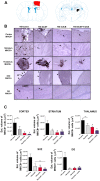
Figure 1. Monotherapy of hUCB or G-CSF, and hUCB+G-CSF combined therapy ameliorate TBI–induced neuroinflammation in gray matter areas.
Downregulation of activated microglial cells in the ipsilateral side of cortical and subcortical gray matter regions after treatment with hUCB alone, G-CSF alone, and combined hUCB+G-CSF relative to saline. Diagram of a coronal section shows the lesion area in red. The squares indicate the region of interest for analyses (Fig. 1A). Photomicrographs of gray matter areas of cortex, striatum, thalamus, SVZ and DG of coronal sections from all four groups (Fig. 1B). Arrows indicate positive staining for activated microglia cells. Quantification of OX-6 immunostaining reflects estimated volume of activated microglia cells of cortex, striatum, thalamus, SVZ, and DG (Fig. 1C). Cortex F3, 20 = 4.913, p<0.0001; striatum F3,20 = 6.466, p<0.0001; thalamus F3,20 = 8.785, p<0.0001; SVZ F3,20 = 6.543, p<0.0001; DG F3,20 = 4.587, p<0.0001. Scale bar in B = 1 µm. * = significant difference between TBI-saline and TBI-G-CSF; & = significant difference between TBI-saline and TBI-hUCB; # = significant difference between TBI-saline and TBI-hADSC; ns = no significance. Significance at p's<0.05.
Figure 2
Figure 2. Monotherapy of hUCB or G-CSF, and hUCB+G-CSF combined therapy ameliorate TBI–induced neuroinflammation in white matter areas.
Downregulation of activated microglia cells in the ipsilateral side of white matter axonal regions after treatment with hUCB alone, G-CSF alone, and combined hUCB+G-CSF relative to TBI-saline. Diagram of a coronal section shows the axonal lesion areas. The squares indicate the region of interest for analyses (Fig. 2A). Photomicrographs of white matter areas: corpus callosum, fornix, and cerebral peduncle of coronal sections from all four groups (Fig. 2B). Arrows indicate positive staining for activated microglia cells. Quantification of OX-6 immunostaining reflects estimated volume of activated microglia cells in corpus callosum, fornix, and cerebral peduncle (Fig. 2C). Corpus callosum, F3,20 = 14.6, p<0.0001; fornix, F3,20 = 9.017, p<0.0001; cerebral peduncle, F3,20 = 4.638, p<0.0001. Scale bar for B = 1 µm. * = significant difference between TBI-saline and TBI-G-CSF; & = significant difference between TBI-saline and TBI-hUCB; # = significant difference between TBI-saline vs TBI-hADSC; ns = no significance. Significance at p's<0.05.
Figure 3
Figure 3. hUCB and G-CSF monotherapy, and combined hUCB+G-CSF attenuate TBI-induced impairment in endogenous neurogenesis and cell loss in rats exposed to chronic TBI.
Significantly enhanced neural differentiation in the DG and SVZ and increased cell survival in CA3 after hUCB or G-CSF monotherapy, and the combined therapy of hUCB+G-CSF relative to saline alone. All 3 treatment conditions percent neurogenesis in the DG (Fig. 3A). Percent neurogenesis in the SVZ (Fig. 3B). Percentage of neuronal survival in the CA3 region of the hippocampus (Fig. 3C). Photomicrographs of SVZ, DG, and CA3 region (Fig. 3D) Top panel arrows indicate positive staining for neurogenesis in SVZ and DG respectivately. Arrow on bottom panel indicates CA3 pyramidal cell loss in TBI-saline (Fig. 3D). F3,20 = 159.3, p<0.0001. Scale bars for Figure D = 50 µm. * = significant difference between TBI-saline and TBI G-CSF; & = significant difference between TBI-saline and TBI-hUCB; # = significantly difference between TBI-saline and TBI-hADSC; ns = no significance. Significance at p's<0.05.
Figure 4
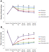
Figure 4. Combined hUCB+G-CSF exert robust functional recovery in chronic TBI.
A separate cohort of TBI animals, using the same experimental paradigm above, was subjected to behavioral tests. Elevated body swing tests revealed that animals displayed normal swing activity (average of 10 swings to both left and right side) prior to TBI (Baseline), but exhibited significant swings to one side after TBI (Day 0 post-TBI) (p's<0.05 versus Baseline) (Fig. 4A). At post-TBI day 7 and day 14, TBI-hUCB+G-GSF and TBI-hUCB alone promoted significantly better recovery compared to G-CSF alone, although all three groups performed better than TBI-saline group (a) (p's<0.05). By day 28, TBI-hUCB and TBI-hUCB+G-CSF were the only two groups that displayed significant recovery of normal swing activity (p's<0.05), while TBI-GCSF group reverted to Day 0 post-TBI levels and did not significantly differ from TBI-saline (Fig. 4A) (p>0.05). By day 56, TBI-hUCB+G-GSF and TBI-hUCB alone were still significantly displaying near normal swing activity (p's<0.05 versus TBI-G-CSF or TBI-saline), but the TBI-hUCB+G-GSF showed significantly better recovery than TBI-hUCB alone (c) (p<0.05). In addition TBI-GCSF group did not significantly differ from TBI-saline by day 56 (Fig. 4A) (p>0.05). Rotorod tests revealed that animals learned to balance on the rotating rod (maximum of 60 seconds) prior to TBI (Baseline), but exhibited significant reduction in balancing time after TBI (Day 0) (p's<0.05 versus Baseline) (Fig. 4B). At post-TBI day 7 and day 14, the performance in balancing on the rotating rod across treatment groups showed the following order of best to least recovery: TBI-hUCB+G-GSF>TBI-hUCB alone>G-CSF alone, with all three groups performing better than TBI-saline group (a) (p's<0.05), but the TBI-hUCB+G-CSF displayed the most effective balancing activity at across all time points (p's<0.05 versus all other groups) (Fig. 4B). By day 28 and day 56, only TBI-hUCB and TBI-hUCB+G-CSF were the only two groups that displayed significant recovery of balancing activity (p's<0.05 versus TBI-G-CSF or TBI-saline), while TBI-GCSF group did not significantly differ from TBI-saline (Fig. 4B) (p's>0.05).

References

    1. Faul M, Xu L, Wald MM, Coronado VG (2010) Traumatic brain injury in the United States: Emergency department visits, hospitalizations and deaths. Atlanta (GA): Centers for Disease Control and Prevention, National Center for Injury Prevention and Control
    1. Fabrizio KS, Keltner NL (2010) Traumatic brain injury in operation enduring freedom/operation iraqi freedom: a primer. The Nursing Clinics of North America 45: 569–580, vi. - PubMed
    1. Ettenhofer KS, Abeles N (2009) The significant of mild traumatic brain injury to cognition and self-reported sympons in long term recovery from injury. J Clin Exp Neuropsychol 31 (3) 363–72. - PubMed
    1. Ozen LJ, Fernandes MA (2012) Slowing down after a mild traumatic brain injury: a strategy to improve cognitive task performance? Archives of clinical neuropsychology : the official journal of the National Academy of Neuropsychologists 27: 85–100. - PubMed
    1. Werner C, Engelhard K (2007) Pathophysiology of traumatic brain injury. British Journal of Anaesthesia 99: 4–9. - PubMed

Publication types

MeSH terms

Substances