. 2014 May;7(3):510-8.
doi: 10.1161/CIRCHEARTFAILURE.113.001023. Epub 2014 Mar 12.

Novel protective role of endogenous cardiac myocyte P2X4 receptors in heart failure

Affiliations

Novel protective role of endogenous cardiac myocyte P2X4 receptors in heart failure

Tiehong Yang et al. Circ Heart Fail. 2014 May.

Abstract

Background: Heart failure (HF), despite continuing progress, remains a leading cause of mortality and morbidity. P2X4 receptors (P2X4R) have emerged as potentially important molecules in regulating cardiac function and as potential targets for HF therapy. Transgenic P2X4R overexpression can protect against HF, but this does not explain the role of native cardiac P2X4R. Our goal is to define the physiological role of endogenous cardiac myocyte P2X4R under basal conditions and during HF induced by myocardial infarction or pressure overload.

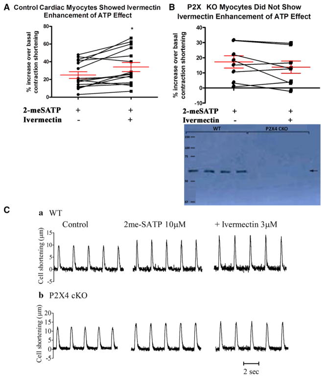
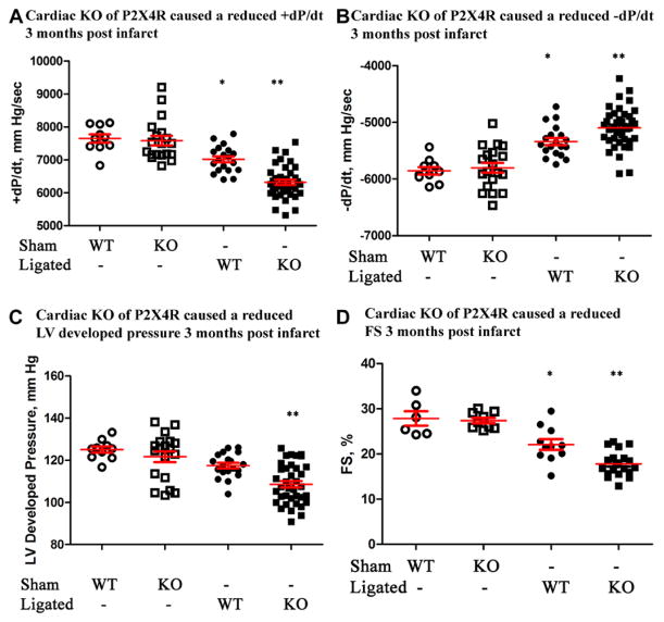
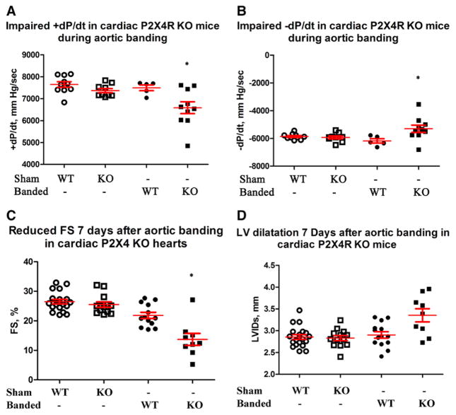
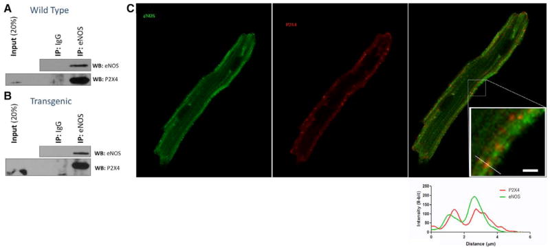
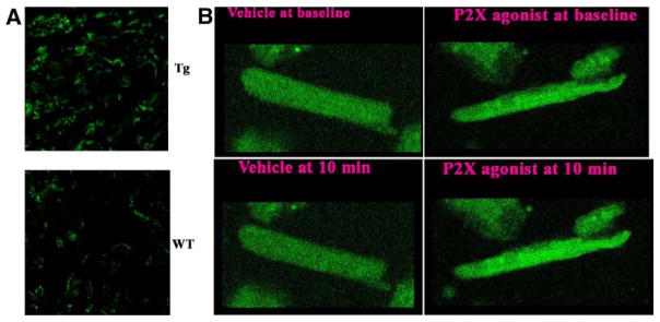
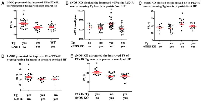
Methods and results: Mice established with conditional cardiac-specific P2X4R knockout were subjected to left anterior descending coronary artery ligation-induced postinfarct or transverse aorta constriction-induced pressure overload HF. Knockout cardiac myocytes did not show P2X4R by immunoblotting or by any response to the P2X4R-specific allosteric enhancer ivermectin. Knockout hearts showed normal basal cardiac function but depressed contractile performance in postinfarct and pressure overload models of HF by in vivo echocardiography and ex vivo isolated working heart parameters. P2X4R coimmunoprecipitated and colocalized with nitric oxide synthase 3 (eNOS) in wild-type cardiac myocytes. Mice with cardiac-specific P2X4R overexpression had increased S-nitrosylation, cyclic GMP, NO formation, and were protected from postinfarct and pressure overload HF. Inhibitor of eNOS, L-N(5)-(1-iminoethyl)ornithine hydrochloride, blocked the salutary effect of cardiac P2X4R overexpression in postinfarct and pressure overload HF as did eNOS knockout.

Conclusions: This study establishes a new protective role for endogenous cardiac myocyte P2X4R in HF and is the first to demonstrate a physical interaction between the myocyte receptor and eNOS, a mediator of HF protection.

Keywords: heart failure; myocytes, cardiac; purines.


Figures

Figure 1
Figure 1
Characterization of heart functions in cardiac-specific knockout (KO) of P2X4 receptors (P2X4R). A, Homozygous P2X4floxed/floxed mice were bred with transgenic mice (Myh6-cre/Esr1*) to generate tamoxifen-inducible cardiac myocyte–specific P2X4R KO. Cardiac ventricular myocytes from control (nonfloxed Myh6-cre/Esr1*) mice (15 cells from 10 mice) showed an increase in contraction shortening in response to P2X agonist 2-methylthioadenosine-5′-triphosphate (2-meSATP; P<0.05 vs basal) and a further increase by the P2X4-specific allosteric enhancer ivermectin (*P<0.05 vs 2-meSATP; repeated-measures ANOVA). B, KO myocytes (8 cells from 6 mice) had a response to 2-meSATP alone (P<0.05 vs basal) but did not show further increase by ivermectin. The percent increase over basal by 2-meSATP alone or by 2-meSATP plus ivermectin was less in KO than in control myocytes (P<0.05). KO myocytes did not have detectable P2X4R by immunoblotting as compared with control myocytes (inset). C, Representative myocyte tracings are shown. Multiple cells within the same mouse were assumed to be independent.
Figure 2
Figure 2
P2X4 receptor (P2X4R) knockout (KO) mice showed a more severe heart failure phenotype after infarction. Wild-type (WT) and KO mice were subjected to sham operation or left anterior descending coronary artery ligation and cardiac functions determined 90 d later. KO hearts had a lower +dP/dt (A), −dP/dt (B), and left ventricular (LV) developed pressure (C) by ex vivo working heart preparation (n=39) as well as a more reduced fractional shortening (FS) by in vivo echocardiography (D; n=20) than WT hearts (n=19 and 11 for ex and in vivo measurements, respectively). Both WT and KO hearts showed lower ±dP/dt and FS than either sham WT (n=10 for ±dP/dt; n=6 for FS) or sham KO (n=19 for ±dP/dt; n=9 for FS) hearts. *P<0.05 ligated WT vs sham WT, sham KO, or ligated KO. **P<0.05 ligated KO vs sham WT, sham KO. P>0.05 sham WT vs sham KO.
Figure 3
Figure 3
P2X4 receptor (P2X4R) knockout (KO) hearts also had a more impaired function after transverse aorta constriction (TAC). Wild-type (WT) and KO mice were subjected to sham operation or TAC and cardiac functions determined 7 d later. KO mice showed a lower +dP/dt (A) and −dP/dt (B) by working heart (n=10), as well as a more reduced fractional shortening (FS; C) and a larger left ventricular internal dimension at systole (LVIDs; D) by echocardiography (n=9) than control WT mice (n=5 for working hearts and n=13 for echocardiography). KO hearts showed lower ±dP/dt, LV developed pressure (LVdevP), and FS compared with either sham WT (n=10 for ±dP/dt and LVdevP; n=19 for FS) or sham KO (n=9 for ±dP/dt and LVdevP; n=13 for FS) hearts. KO hearts also showed more dilated LVIDs compared with sham WT or sham KO hearts. *P<0.05 banded KO vs banded WT, sham WT, or sham KO; P>0.05 sham WT vs KO; P>0.05 banded WT vs sham WT or sham KO in ±dP/dt and LVIDs; P<0.05 banded WT vs sham WT or sham KO in FS comparison.
Figure 4
Figure 4
Endothelial nitric oxide synthase (eNOS) and P2X4 receptor (P2X4R) coimmunoprecipitated in cardiac ventricular myocytes of wild-type (WT) and P2X4-Tg hearts. A, WT myocyte lysates were incubated overnight with anti-eNOS antibody or with nonspecific IgG as control. The isolated complex was subjected to Western blot (WB) analysis and probed with eNOS (top) and P2X4 (bottom) antibodies. IP indicates immunoprecipitation; and Tg, transgenic. As a control, eNOS coimmunoprecipitated itself. Coimmunoprecipitation of P2X4R with eNOS antibody (lane 3) but not with control IgG (lane 2) is shown. B, Same experiment as in A conducted in P2X4-Tg myocytes. C, Immunostaining of eNOS (green), P2X4R (red), and merged image is shown for a P2X4R-overexpressing Tg cardiac myocyte.
Figure 5
Figure 5
Increased cardiac protein S-nitrosylation levels and nitric oxide formation in P2X4 receptor (P2X4R) transgenic (Tg) animals. In A, a typical example of S-nitrosylated proteins in P2X4R-overexpressing Tg and wild-type (WT) hearts (n=5 for each) is shown as determined using biotin switch method. The counts of total summed intensities produced 1 distribution for each heart and analyzed with Kolmogorov–Smirnov test for equality of distributions. The test is rejected (P<0.001) in favor of the distributions not being the same. B, Typical changes in DAF-FM intensities at baseline vs after exposure to agonist or vehicle. Note the declined intensity in vehicle-treated cells due to photobleaching of fluorescein.
Figure 6
Figure 6
Pharmacological inhibition by L-N-(1-iminoethyl)ornithine hydrochloride (L-NIO) and genetic knockout (KO) of endothelial NO synthase (eNOS) abrogated the improved cardiac function by P2X4 receptor (P2X4R) overexpression in heart failure. A, Daily injection of L-NIO 3 d before and 7 d after left anterior descending coronary artery (LAD) ligation in P2X4R-overexpressing transgenic (Tg) mice (n=13) resulted in a reduced fractional shortening (FS) compared with Tg mice not receiving drug (ie, receiving vehicle n=24). Tg mice receiving L-NIO had similar FS as WT mice (n=21 WT) receiving the drug. KO of eNOS in P2X4R Tg mice blocked the improved +dP/dt (B) and FS (C) by P2X4R overexpression in postinfarction heart failure. Thirty days after LAD ligation, P2X4R Tg mice (n=20) showed better +dP/dt compared with P2X4R Tg/eNOS KO (n=22), non-Tg WT (n=9), or eNOS KO mice (n=21). Similarly, P2X4R Tg mice (n=9) had greater FS than any other genotype at 30 d after ligation. Non-Tg WT (n=10), eNOS KO (n=14), P2X4R Tg/eNOS KO mice (n=12) did not differ in FS (ANOVA and post-test comparison). D, Using the same drug injection protocol as in A, P2X4R Tg mice treated with vehicle subjected to transverse aorta constriction (TAC; n=22) had higher FS compared with Tg animals that had received L-NIO (n=11); WT animals (n=11) and Tg mice treated with L-NIO had similar FS. E, 21 days after TAC, P2X4R Tg mice (n=8) showed better FS compared with P2X4R Tg/eNOS KO (n=12) or eNOS KO mice (n= 13). *P<0.05 vs any other group. P>0.05 among groups not marked*.

References

    1. Fredholm BB, IJzerman AP, Jacobson KA, Linden J, Müller C. Nomenclature and classification of adenosine receptors – an update. Pharmacol Rev. 2011;63:1–34. - PMC - PubMed
    1. Oh EY, Abraham T, Saad N, Rapp JH, Vastey FL, Balmir E. A comprehensive comparative review of adenosine diphosphate receptor antagonists. Expert Opin Pharmacother. 2012;13:175–191. - PubMed
    1. Fredholm BB, Abbracchio MP, Burnstock G, Daly JW, Harden TK, Jacobson KA, Leff P, Williams M. Nomenclature and classification of purinoceptors. Pharmacol Rev. 1994;46:143–156. - PMC - PubMed
    1. Khakh BS, Burnstock G. The double life of ATP. Sci Am. 2009;301:84–90. 92. - PMC - PubMed
    1. Yang R, Liang BT. Cardiac P2X4 receptors: targets in ischemia and HF? Circ Res. 2012;111:397–401. - PubMed

Publication types

Substances