. 2014 Mar 12;9(3):e91466.
doi: 10.1371/journal.pone.0091466. eCollection 2014.

Transcriptome profiling of a multiple recurrent muscle-invasive urothelial carcinoma of the bladder by deep sequencing

Affiliations

Transcriptome profiling of a multiple recurrent muscle-invasive urothelial carcinoma of the bladder by deep sequencing

Shufang Zhang et al. PLoS One. .

Abstract

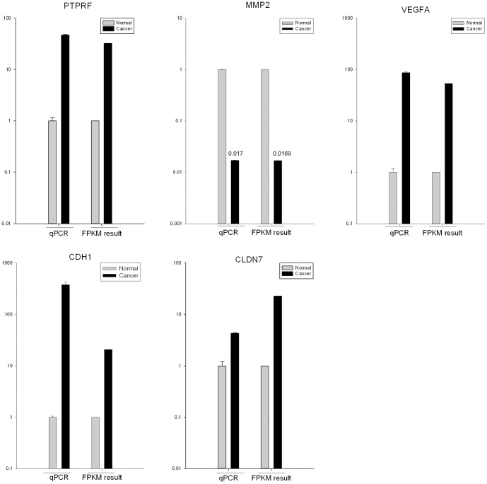
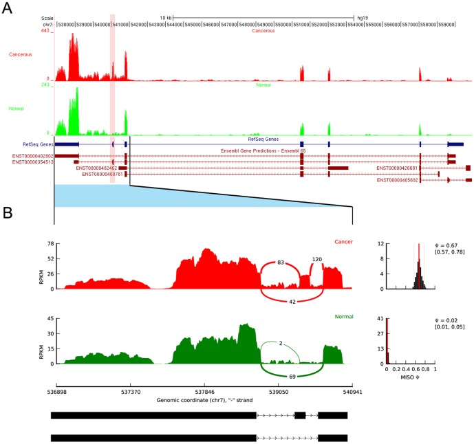
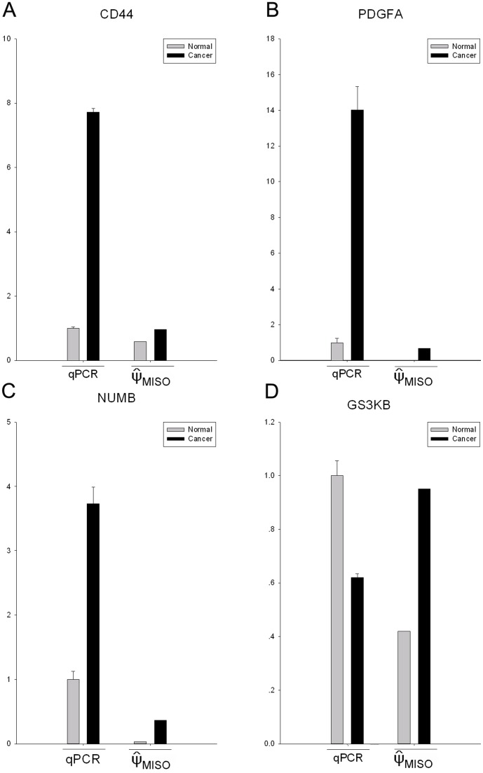
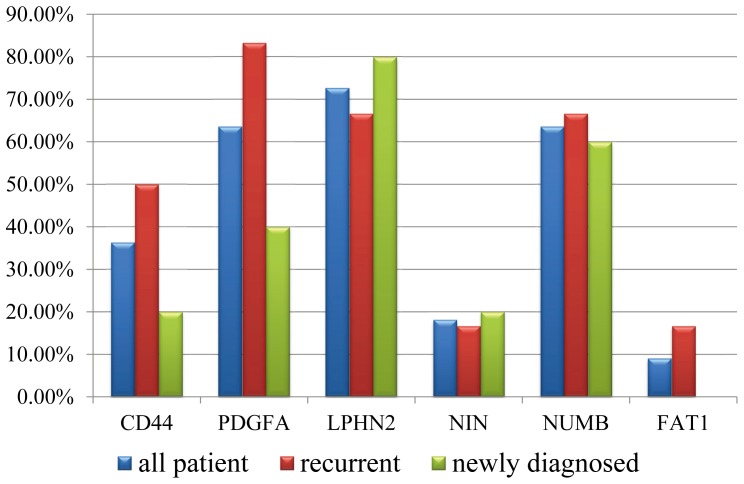
Urothelial carcinoma of the bladder (UCB) is one of the commonly diagnosed cancers in the world. The UCB has the highest rate of recurrence of any malignancy. A genome-wide screening of transcriptome dysregulation between cancer and normal tissue would provide insight into the molecular basis of UCB recurrence and is a key step to discovering biomarkers for diagnosis and therapeutic targets. Compared with microarray technology, which is commonly used to identify expression level changes, the recently developed RNA-seq technique has the ability to detect other abnormal regulations in the cancer transcriptome, such as alternative splicing. In this study, we performed high-throughput transcriptome sequencing at ∼50× coverage on a recurrent muscle-invasive cisplatin-resistance UCB tissue and the adjacent non-tumor tissue. The results revealed cancer-specific differentially expressed genes between the tumor and non-tumor tissue enriched in the cell adhesion molecules, focal adhesion and ECM-receptor interaction pathway. Five dysregulated genes, including CDH1, VEGFA, PTPRF, CLDN7, and MMP2 were confirmed by Real time qPCR in the sequencing samples and the additional eleven samples. Our data revealed that more than three hundred genes showed differential splicing patterns between tumor tissue and non-tumor tissue. Among these genes, we filtered 24 cancer-associated alternative splicing genes with differential exon usage. The findings from RNA-Seq were validated by Real time qPCR for CD44, PDGFA, NUMB, and LPHN2. This study provides a comprehensive survey of the UCB transcriptome, which provides better insight into the complexity of regulatory changes during recurrence and metastasis.


Conflict of interest statement

Competing Interests: The authors have declared that no competing interests exist. Yongqing Ye is employed by the Department of Shanghai Claison Bio-Technology, which was involved in helping the authors performthe sample collection. The authors have no other relationship with the Department of Shanghai Claison Bio-Technology relating to employment, consultancy, patents, products in development or marketed products. This does not alter the authors'? adherence to all the PLOS ONE policies on sharing data and materials.

Figures

Figure 1
Figure 1. The differentially expressed genes detected by RNA-seq are confirmed by qRT-PCR.
qRT-PCR was performed for five genes that are identified as differential expressed genes between UCB and non-tumor tissues. The expression level of each gene was normalized to the level in non-tumor tissue. A-E: PTPRF, MMP2, VEGFA, CDH1 and CLDN7.
Figure 2
Figure 2. qRT-PCR validation of the differentially expressed genes in the additional patients.
qRT-PCR was performed for five differentially expressed genes (CDH1, VEGFA, PTPRF, CLDN7 and MMP2) in the additional 11 patients (including 6 recurrent and drug-resistant UCB patients and 5 newly diagnosed patients). The histogram showed the proportion of validated patients in all cases (blue), the recurrent cases (red) and newly diagnosed (green).
Figure 3
Figure 3. RNA-Seq read mapping to the reference gene PDGFA.
A: RNA-Seq read mapping to the UCSC reference genome (hg19) of the gene PDGFA for UCB and non-tumor tissues in this study. The UCB tracks are shown in red and non-tumor tissue in green. The pink band indicated the location of skipped exon. B: The detail of junction reads mapping to the skipped exon and its neighboring exons. The Ψ (”percentage spliced in”) indicates the ratio of reads supporting inclusion exon vs. total reads supporting both inclusion and exclusion exon. The Ψ posterior distributions were shown in the right side.
Figure 4
Figure 4. The qRT-PCR validation of differential splicing events detected by RNA-seq.
qRT-PCR was performed for four genes that are identified as differential splicing genes between UCB and non-tumor tissues. The result of qRT-PCR is the relative expression level of the skipped exon and the neighboring constitutive exon. The expression level of each exon was normalized to the level in non-tumor tissue. The ΨMISO was the result of MISO, indicates the ratio of reads supporting inclusion exon vs. total reads supporting both inclusion and exclusion exon. A∼D: CD44, PDGFA, NUMB and GSK3B.
Figure 5
Figure 5. qRT-PCR validation of differential splicing events in the additional patients.
qRT-PCR was performed for six differential splicing genes (CD44, PDGFA, NUMB, LPHN2, NIN and FAT1) in the additional 11 patients (including 6 recurrent and drug-resistant UCB patients and 5 newly diagnosed patients). The histogram showed the proportion of validated patients in all cases (blue), the recurrent cases (red) and newly diagnosed (green).

References

    1. Jemal A, Siegel R, Ward E, Hao Y, Xu J, et al. (2008) Cancer statistics, 2008. CA Cancer J Clin 58: 71–96. - PubMed
    1. Botteman MF, Pashos CL, Redaelli A, Laskin B, Hauser R (2003) The health economics of bladder cancer: a comprehensive review of the published literature. Pharmacoeconomics 21: 1315–1330. - PubMed
    1. Hayne D, Arya M, Quinn MJ, Babb PJ, Beacock CJ, et al. (2004) Current trends in bladder cancer in England and Wales. J Urol 172: 1051–1055. - PubMed
    1. Knowles MA (2001) What we could do now: molecular pathology of bladder cancer. Molecular Pathology 54: 215–221. - PMC - PubMed
    1. Williams SG, Stein JP (2004) Molecular pathways in bladder cancer. Urological Research 32: 373–385. - PubMed

Publication types

MeSH terms

Associated data