. 2014 Mar 12;6(227):227ra34.
doi: 10.1126/scitranslmed.3006927.

TGF-β signaling mediates endothelial-to-mesenchymal transition (EndMT) during vein graft remodeling

Affiliations

TGF-β signaling mediates endothelial-to-mesenchymal transition (EndMT) during vein graft remodeling

Brian C Cooley et al. Sci Transl Med. .

Abstract

Veins grafted into an arterial environment undergo a complex vascular remodeling process. Pathologic vascular remodeling often results in stenosed or occluded conduit grafts. Understanding this complex process is important for improving the outcome of patients with coronary and peripheral artery disease undergoing surgical revascularization. Using in vivo murine cell lineage-tracing models, we show that endothelial-derived cells contribute to neointimal formation through endothelial-to-mesenchymal transition (EndMT), which is dependent on early activation of the Smad2/3-Slug signaling pathway. Antagonism of transforming growth factor-β (TGF-β) signaling by TGF-β neutralizing antibody, short hairpin RNA-mediated Smad3 or Smad2 knockdown, Smad3 haploinsufficiency, or endothelial cell-specific Smad2 deletion resulted in decreased EndMT and less neointimal formation compared to controls. Histological examination of postmortem human vein graft tissue corroborated the changes observed in our mouse vein graft model, suggesting that EndMT is operative during human vein graft remodeling. These data establish that EndMT is an important mechanism underlying neointimal formation in interpositional vein grafts, and identifies the TGF-β-Smad2/3-Slug signaling pathway as a potential therapeutic target to prevent clinical vein graft stenosis.


Figures

Fig. 1
Fig. 1. Endothelial cell lineage tracing during vein graft remodeling
(A) Endothelial YFP expression in ungrafted (uninjured) jugular veins from EndotrackYFP and Ind.EndotrackYFP mice as well as in grafted veins from recombined and non-recombined EndotrackYFP and Ind.EndotrackYFP mice grafted into wild type recipients at 35 days. Scale bars, 30 μm (uninjured) and 100 μm. (B) The number of YFP+ cells per neointimal cells was quantified. Data are means ± SEM (n=5). (C) Endothelial-derived (YFP+) and non–endothelial-derived (YFP) cells in the neointima over time after engraftment. Data are means ± SEM (n=4). (D) BrdU+ cells relative to the total number of neointimal cells over time after engraftment. Data are means ± SEM (n=4). P-values in (B to D) determined by Student's t test.
Fig. 2
Fig. 2. EndMT during vein graft remodeling
EndotrackYFP veins were grafted into wild type recipients (n = 5) and analyzed over the course of 35 days. (A) Immunofluorescence staining of the endothelial marker CD31 in grafted veins. (B) The proportion of YFP+ cells expressing CD31, VE-cadherin, and endoglin over time. (C) SMA expression by YFP+ cells. (D) The percentage of YFP+ cells expressing immature VSMC markers SMA and SM22α and mature VSMC markers smoothelin, calponin, and SM-MHC. (E) SM-MHC expression in non– endothelial-derived (YFP) neointimal cells. (F) The percentage of non–endothelial-derived (YFP) neointimal cells expressing both immature and mature VSMC markers. (G) Expression of CD31 and SMA in endothelial lineage–derived (YFP+) cells 7 days after grafting. Scale bars in (A, C, E, and G), 10 μm. L, lumen. Data in (B, D, and F) are means ± SEM (n=5). P-values determined by Student's t test. *P< 0.05, **P<0.01, ***P < 0.001
Fig. 3
Fig. 3. TGF-β signaling during EndMT in vivo
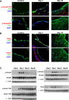
EndotrackYFP veins were grafted into wild type recipients (n = 3). (A) Immunofluorescence staining of phosphorylated Smad1/5/8 and Smad2/3 after grafting in both endothelial-derived (YFP+) and non–endothelial-derived (YFP) neointimal cells. Scale bars, 5 μm. L, lumen. (B) Immunostaining of the TGF-β–regulated transcription factors Slug and Twist in endothelial-derived cells after grafting. Scale bars, 10 μm. L, lumen. (C) Western blot analysis of p-Smad3, p-Smad2, p-Smad1/5/8, and Slug from day 0 to 35 after grafting. β-actin served as loading control.
Fig. 4
Fig. 4. A TGF-β neutralizing antibody reduces TGF-β signaling and EndMT in vivo
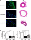
EndotrackYFP veins were immersed in TGF-β neutralizing antibody or control IgG for 4 hours prior to grafting. TGF-β signaling was blocked in recipient wild-type animals by treatment with a pan–TGF-β neutralizing antibody prior to grafting, and every 14 days thereafter (n = 5 grafts per group). (A) Plasma levels of TGF-β1 after vein graft. Data are means ± SEM (n=5). **P<0.01, ***P < 0.001, ****P < 0.0001 ANOVA with Newman-Keuls Multiple Comparison test. (B) YFP expression and corresponding H&E histology of vein grafts from treated mice at day 35. Scale bars, 15 μm in YFP images, 100 μm in H&E images. L, lumen. (C) Percentage of endothelial-derived (YPF+) cells in neointima of vein grafts treated with control or TGF-β neutralizing antibody. (D) Quantification of neointimal area in treated vein grafts harvested at day 35 after grafting. (E) The percentage of SM22α+ and SM-MHC+ cells in the neointima at day 35. Data are means ± SEM (n=5). P-values in (C to E) determined by Student's t test.
Fig. 5
Fig. 5. Knockdown of Smad3 or Smad2 attenuates EndMT in mouse vein grafts
EndotrackYFP veins transduced with Smad3 or Smad2 shRNA were grafted into wild type recipients (n = 5). (A) Immunofluorescence staining and H&E histology of the vein grafts harvested at day 35. Scale bars, 10 μm in YFP images, 100 μm in H&E images. L, lumen. (B) Quantification of the neointimal area and endothelial-derived (YFP+) cells after knockdown of Smad3 or Smad2. Data are means ± SEM (n = 5). P values determined by Student's t test.
Fig. 6
Fig. 6. Neointimal formation and EndMT in vein grafts is regulated via Smad3 and Slugsignaling
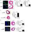
All data were obtained at day 35 post-grafting. (A) YFP and H&E staining of Smad3+/- EndotrackYFP veins grafted into wild type recipients. Scale bars, 10 μm for YFP images; 100 μm for H&E images. L, lumen. (B) Quantification of neointima area and endothelial-derived (YFP+) cells in the neointima. (C) H&E staining and quantification of neointima in Smad3+/- mouse veins overexpressing Slug that had been grafted into wild type recipients (n = 5). (D) Vein grafts from Ind.EndotrackYFP and Smad2Fl/Fl;Ind.EndotrackYFP mice that had been transplanted into wild type recipients (n = 5) were stained for YFP or H&E. Scale bars, 15 μm for YFP images L, lumen; 100 μm for H&E images. Quantification of YFP+ endothelial-derived cells and neointimal area. Data in (B to D) are means ± SEM (n = 5). P-values determined by Student's t test.
Fig. 7
Fig. 7. EndMT in human endothelial cells and in human vein graft remodeling
(A) HUVECs were treated with 10 ng/ml TGF-β1 and stained for CD31, VE-cadherin, SM22α, Slug, and SM-MHC. Scale bars, 10 μm. Images are representative of n=3 experiments. (B) Human RT2 PCR Arrays were used to assess gene expression in baseline HUVECs ± TGF-β1 and human aortic VSMCs. Data represent significant (P≤0.05, Student's t test) 2-fold differentially regulated gene expression in HUVECs compared to HUVECs with TGF-β1. (C) EndMT observed in tissue samples after during human vein graft remodeling. Immunofluorescence co-staining of early (<1 year, n=5) and late (>6 year, n=5) human vein grafts with endothelial (VE-cadherin, vWF) and immature (SMA, SM22α) or mature (SM-MHC, calponin) VSMC markers. Scale bars, 100 μm. Graph depicts quantification of EndMT (double-positive staining) in human vein grafts (n=5 per group). P-values determined by Student's t test.

Comment in

References

    1. Roger VL, Go AS, Lloyd-Jones DM, Adams RJ, Berry JD, Brown TM, Carnethon MR, Dai S, de Simone G, Ford ES, Fox CS, Fullerton HJ, Gillespie C, Greenlund KJ, Hailpern SM, Heit JA, Ho PM, Howard VJ, Kissela BM, Kittner SJ, Lackland DT, Lichtman JH, Lisabeth LD, Makuc DM, Marcus GM, Marelli A, Matchar DB, McDermott MM, Meigs JB, Moy CS, Mozaffarian D, Mussolino ME, Nichol G, Paynter NP, Rosamond WD, Sorlie PD, Stafford RS, Turan TN, Turner MB, Wong ND, Wylie-Rosett J. Heart disease and stroke statistics--2011 update: a report from the American Heart Association. Circulation. 2011 Feb 1;123:e18. - PMC - PubMed
    1. Mehta RH, Ferguson TB, Lopes RD, Hafley GE, Mack MJ, Kouchoukos NT, Gibson CM, Harrington RA, Califf RM, Peterson ED, Alexander JH. Saphenous Vein Grafts With Multiple Versus Single Distal Targets in Patients Undergoing Coronary Artery Bypass Surgery: One-Year Graft Failure and Five-Year Outcomes From the Project of Ex-Vivo Vein Graft Engineering via Transfection (PREVENT) IV Trial. Circulation. 2011 Jul 19;124:280. - PMC - PubMed
    1. Mitra AK, Gangahar DM, Agrawal DK. Cellular, molecular and immunological mechanisms in the pathophysiology of vein graft intimal hyperplasia. Immunol Cell Biol. 2006 Apr;84:115. - PubMed
    1. Boehm M, Olive M, True AL, Crook MF, San H, Qu X, Nabel EG. Bone marrow-derived immune cells regulate vascular disease through a p27(Kip1)-dependent mechanism. J Clin Invest. 2004 Aug;114:419. - PMC - PubMed
    1. Olive M, Mellad JA, Beltran LE, Ma M, Cimato T, Noguchi AC, San H, Childs R, Kovacic JC, Boehm M. p21Cip1 modulates arterial wound repair through the stromal cell-derived factor-1/CXCR4 axis in mice. J Clin Invest. 2008 Jun;118:2050. - PMC - PubMed

Publication types

MeSH terms