

Review
doi: 10.1128/JVI.03404-13.
Epub 2014 Mar 12.
Dengue virus- and hepatitis C virus-induced replication and assembly compartments: the enemy inside--caught in the web
Affiliations
- PMID: 24623440
- PMCID: PMC4093888
- DOI: 10.1128/JVI.03404-13
Item in Clipboard
Review
Dengue virus- and hepatitis C virus-induced replication and assembly compartments: the enemy inside--caught in the web
J Virol.
2014 Jun.
Abstract
Dengue virus (DENV) and hepatitis C virus (HCV), members of the family Flaviviridae, are global human health concerns. As positive-strand RNA viruses, they each replicate in the cytoplasm of infected cells and induce distinct membranous replication compartments where most, if not all, steps of the viral life cycle occur. This Gem briefly reviews the most recent insights into the architecture and functional properties of membranous replication and assembly sites induced by DENV and HCV.
Figures
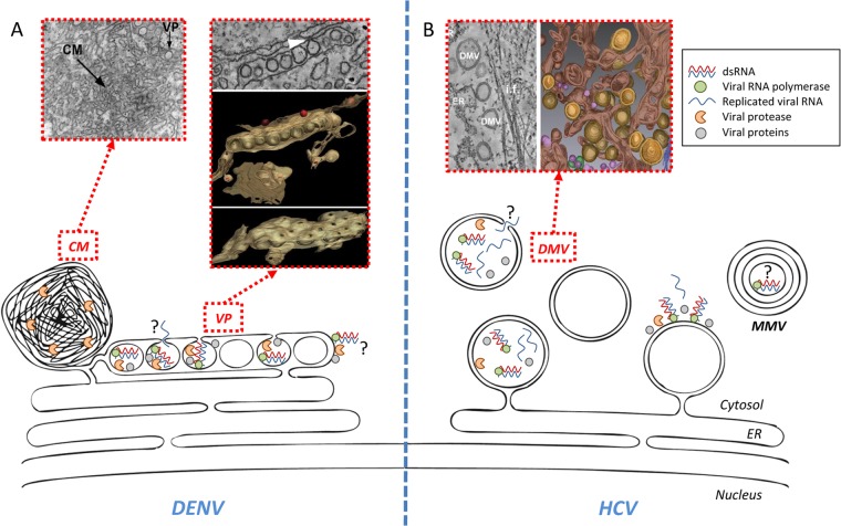
DENV and HCV replication factories and associated membranes. (A) Schematic representation of DENV-induced endoplasmic reticulum (ER)-derived vesicle packets (VP) and convoluted membranes (CM). Corresponding transmission EM pictures (for VP and CM) and a 3D reconstruction from electron tomography (for VP) are also shown. The identified pore of each vesicle might act as an exit site for neosynthesized viral RNA. Note that DENV RNA replication on the cytosolic side of VPs is not excluded. (B) Schematic representation of HCV-induced membrane rearrangements. Note that the appearance of double-membrane vesicles (DMVs) coincides with the peak of RNA replication. At later time points after infection, multimembrane vesicles (MMV) as well as double-membrane tubes (not shown) are also observed, but their involvement in RNA replication remains to be determined. An opening toward the cytoplasm is observed in ∼10% of DMVs and might allow exit of viral RNA. Although enzymatically active HCV replicase is physically associated with DMVs, it is not known if RNA amplification occurs within DMVs and/or on their cytosolic side. Some of the insets are reprinted from reference with permission from the publisher and from PLoS Pathogens (3).
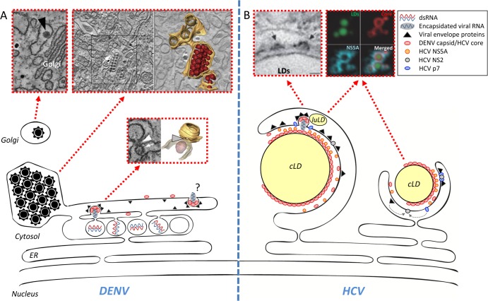
DENV and HCV assembly sites. (A) Schematic model of DENV assembly. Three populations of virions are observed in the EM: first, budding viruses next to the pores of VPs (electron densities of detected structures presumably correspond to RNA); second, virus stacks within the lumen of the VP-containing ER network; third, viruses in Golgi-related vesicles. Note that virus budding might also occur in other areas of the ER and remote from VPs. Transmission EM images and 3D reconstructions of an electron tomogram are shown above the schematic. (B) Schematic model of HCV assembly. The majority of core protein is located on the surface of cytoplasmic lipid droplets (cLD), where a fraction of NS5A also resides. NS2 is thought to orchestrate the assembly process by recruiting on one hand p7, along with the two envelope glycoproteins, and on the other hand NS3 (not shown), eventually along with NS5A or the complete viral replicase (NS3 to NS5B). P7 is important for the envelopment of assembled capsids containing the HCV RNA genome. Assembly also involves components of the LDL synthesis machinery, including luminal lipid droplets (luLD) and apolipoproteins, most notably ApoE (not shown). Some of the insets are reprinted from reference with permission from the publisher and from Nature Cell Biology (15).
References
-
- Romero-Brey I, Merz A, Chiramel A, Lee JY, Chlanda P, Haselman U, Santarella-Mellwig R, Habermann A, Hoppe S, Kallis S, Walther P, Antony C, Krijnse-Locker J, Bartenschlager R. 2012. Three-dimensional architecture and biogenesis of membrane structures associated with hepatitis C virus replication. PLoS Pathog. 8:e1003056. 10.1371/journal.ppat.1003056 - DOI - PMC - PubMed
-
- Welsch S, Miller S, Romero-Brey I, Merz A, Bleck CK, Walther P, Fuller SD, Antony C, Krijnse-Locker J, Bartenschlager R. 2009. Composition and three-dimensional architecture of the dengue virus replication and assembly sites. Cell Host Microbe 5:365–375. 10.1016/j.chom.2009.03.007 - DOI - PMC - PubMed
Publication types
MeSH terms
LinkOut - more resources
Full Text Sources
Other Literature Sources
Research Materials
