. 2014 Mar 12;34(11):3901-9.
doi: 10.1523/JNEUROSCI.4070-13.2014.

CCR2+ Ly6C(hi) inflammatory monocyte recruitment exacerbates acute disability following intracerebral hemorrhage

Affiliations

CCR2+ Ly6C(hi) inflammatory monocyte recruitment exacerbates acute disability following intracerebral hemorrhage

Matthew D Hammond et al. J Neurosci. .

Abstract

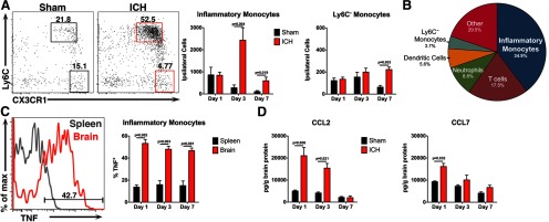
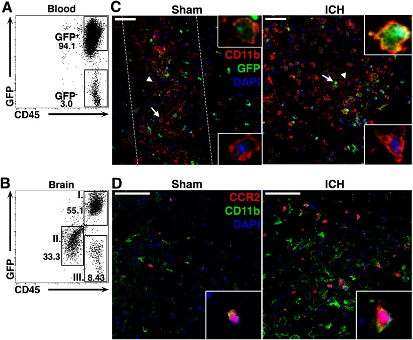
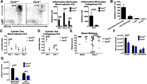
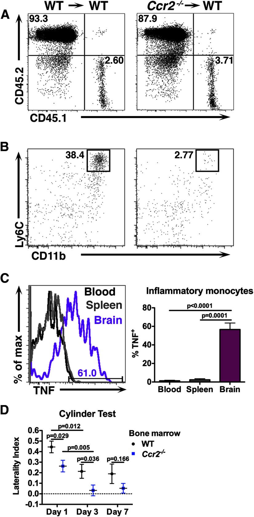
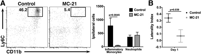
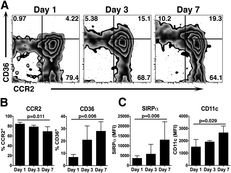
Intracerebral hemorrhage (ICH) is a devastating type of stroke that lacks a specific treatment. An intense immune response develops after ICH, which contributes to neuronal injury, disability, and death. However, the specific mediators of inflammation-induced injury remain unclear. The objective of the present study was to determine whether blood-derived CCR2+ Ly6C(hi) inflammatory monocytes contribute to disability. ICH was induced in mice and the resulting inflammatory response was quantified using flow cytometry, confocal microscopy, and neurobehavioral testing. Importantly, blood-derived monocytes were distinguished from resident microglia by differential CD45 staining and by using bone marrow chimeras with fluorescent leukocytes. After ICH, blood-derived CCR2+ Ly6C(hi) inflammatory monocytes trafficked into the brain, outnumbered other leukocytes, and produced tumor necrosis factor. Ccr2(-/-) mice, which have few circulating inflammatory monocytes, exhibited better motor function following ICH than control mice. Chimeric mice with wild-type CNS cells and Ccr2(-/-) hematopoietic cells also exhibited early improvement in motor function, as did wild-type mice after inflammatory monocyte depletion. These findings suggest that blood-derived inflammatory monocytes contribute to acute neurological disability. To determine the translational relevance of our experimental findings, we examined CCL2, the principle ligand for the CCR2 receptor, in ICH patients. Serum samples from 85 patients were collected prospectively at two hospitals. In patients, higher CCL2 levels at 24 h were independently associated with poor functional outcome at day 7 after adjusting for potential confounding variables. Together, these findings suggest that inflammatory monocytes worsen early disability after murine ICH and may represent a therapeutic target for patients.

Keywords: CCL2; CCR2; bone marrow chimeras; intracerebral hemorrhage; monocytes; neuroinflammation.


Figures

Figure 1.
Figure 1.
Inflammation following ICH is characterized by a monocyte-rich cellular infiltrate. Mice were subjected to sham or ICH and their brains were analyzed to characterize the cellular and molecular inflammatory composition. A, Representative flow cytometry plots from day 3 show that inflammatory monocytes (Ly6Chi CX3CR1low) outnumber Ly6C monocytes (Ly6C CX3CR1hi) in ICH-treated brains from Cx3cr1GFP/+ mice, whereas both populations are low in number in sham brains. Numbers in the plots indicate the percentage of cells in the plot located in each respective gate. Plots are gated on CD45hi, CD3, Ly6G. N = 5–10 mice per group. B, This pie chart shows the mean of each blood-derived cell population in Cx3cr1GFP/+ ICH brains at day 3. Each population was calculated as a percentage of total CD45hi cells in the ipsilateral brain hemisphere. N = 9. C, This representative histogram from WT brain and spleen 3 d after ICH shows inflammatory monocyte TNF staining. Roughly half of the inflammatory monocytes isolated from ICH brains were positive for TNF up to day 7, whereas significantly fewer inflammatory monocytes from spleens stained positive for TNF. N = 5–9 per group. D, By ELISA, the monocyte chemoattractant CCL2 is increased in WT ICH brains up to 3 d after ICH, whereas CCL7 is increased only at day 1. N = 4–6 per group. Error bars indicate SEM.
Figure 2.
Figure 2.
Blood-derived monocytes are abundant in the brain after ICH. A, WT mice transplanted with BM from a mouse constitutively expressing GFP were used to distinguish between resident microglia and blood-derived leukocytes. At least 8 weeks after transplantation, the vast majority of leukocytes (CD45+ cells) were found to originate from GFP donor cells. B, Three days following ICH, the majority of CD45hi peripheral leukocytes in the brain were GFP+ (population I), rather than GFP (population III). Microglia were GFP, CD45low (population II). C, Staining for CD11b (red), GFP (green), and DAPI (blue) shows that many of the globoid CD11b+ cells within the hematoma are also GFP+ (arrows and upper insets), indicating their BM origin. Some GFP microglia can also be seen (arrowheads and lower insets). Dotted lines in sham indicate localized inflammation due to injury from the needle insertion. D, Ccr2RFP/+ mice 3 d following injury have CD11b+ (green) and CCR2+ (red) inflammatory monocytes present around the hematoma. DAPI is shown in blue. Scale bars: 50 μm.
Figure 3.
Figure 3.
Ccr2−/− mice exhibit decreased inflammatory monocyte recruitment and are protected from early motor deficits. Because Ccr2−/− mice have few circulating inflammatory monocytes, they were used to evaluate the functional and inflammatory effects of monocytes following ICH. A, Inflammatory monocytes are significantly less abundant in Ccr2−/− brains following either blood injection ICH or collagenase ICH. Plots are representative from day 3 blood injection ICH and are gated on CD45hi, CD3, Ly6G. N = 7–9 blood injection ICH; n = 3–4 collagenase ICH. B, The majority of CCR2+ cells in ipsilateral control brains are inflammatory monocytes. N = 3–9. C, One day following blood injection ICH, Ccr2−/− mice display a decreased left forelimb deficit. N = 9–15 per group. D, In the collagenase ICH model, Ccr2−/− mice also display a decreased forelimb deficit at day 1. N = 3–5. E, Three days following blood injection ICH Ccr2−/− mice perform better on the beam balance test, indicating that mice with fewer inflammatory monocytes have abrogated secondary injury. Horizontal lines indicate median and bars show interquartile range. N = 8–14. F, Ccr2−/− mice have similar lymphocyte and neutrophil numbers in blood compared with littermate controls. N = 11–17. G, Ccr2−/− mice have many fewer inflammatory monocytes and slightly fewer Ly6C monocytes in blood. N = 13–17. Error bars in AD, F, and G indicate SEM.
Figure 4.
Figure 4.
Blood-derived inflammatory monocytes contribute to secondary disability after ICH. WT CD45.1 mice were lethally irradiated, transplanted with Ccr2−/− BM, and subjected to ICH at least 8 weeks later. A, The majority of leukocytes in the blood of chimeric mice in both genotypes were derived from donor BM. B, Mice given Ccr2−/− BM were found to have fewer inflammatory monocytes in their brains as early as 12 h following ICH. Plots are gated on CD45hi, CD3, Ly6G. C, This representative histogram from a chimeric mouse with WT BM shows that inflammatory monocytes isolated from brain 3 d after ICH make TNF, but those found in blood and spleen do not make appreciable levels. N = 5. D, Chimeric mice given Ccr2−/− BM display a decreased left forelimb deficit 1 and 3 d following ICH, suggesting that blood-derived inflammatory monocytes are responsible for inflicting secondary injury following ICH. Both groups improve from day 1 to 3. N = 8–18. Error bars indicate SEM.
Figure 5.
Figure 5.
MC-21 treatment diminishes the inflammatory monocyte response to ICH. A, WT mice were treated with the anti-CCR2 depletion antibody MC-21 before and after ICH. Fewer CD11b+, Ly6Chi inflammatory monocytes were detected in the brains of MC-21-treated mice, but no differences were seen in neutrophils. The plots are gated on CD45hi, CD3, Ly6G cells. N = 5–7. B, Mice treated with MC-21 display a significantly reduced left forelimb deficit 1 d after ICH. N = 7–8. Error bars indicate SEM.
Figure 6.
Figure 6.
Inflammatory monocytes isolated from ICH brains change phenotype over time. WT mice were subjected to ICH and brains were analyzed by flow cytometry. A, These plots are gated on CD45hi, CD3, Ly6G, Ly6Chi inflammatory monocytes isolated at the indicated time points. CCR2 decreases as CD36 increases between day 1 and 7. Numbers indicate the percentage of total cells on each plot represented in each respective quadrant. B, Quantification from the data shown in A indicates that the percentage of CCR2+ inflammatory monocytes decreases as the percentage of CD36+ inflammatory monocytes increases. Graphs depict mean ± SD. N = 3–6. C, The mean fluorescence intensity (MFI) of SIRPα and CD11c on inflammatory monocytes increases from day 1 to 7. Graphs depict mean ± SD. N = 3–6.

References

    1. Agnihotri S, Czap A, Staff I, Fortunato G, McCullough LD. Peripheral leukocyte counts and outcomes after intracerebral hemorrhage. J Neuroinflammation. 2011;8:160. doi: 10.1186/1742-2094-8-160. - DOI - PMC - PubMed
    1. Azcutia V, Stefanidakis M, Tsuboi N, Mayadas T, Croce KJ, Fukuda D, Aikawa M, Newton G, Luscinskas FW. Endothelial CD47 promotes vascular endothelial-cadherin tyrosine phosphorylation and participates in T cell recruitment at sites of inflammation in vivo. J Immunol. 2012;189:2553–2562. doi: 10.4049/jimmunol.1103606. - DOI - PMC - PubMed
    1. Banks JL, Marotta CA. Outcomes validity and reliability of the modified Rankin scale: implications for stroke clinical trials: a literature review and synthesis. Stroke. 2007;38:1091–1096. doi: 10.1161/01.STR.0000258355.23810.c6. - DOI - PubMed
    1. Boring L, Gosling J, Chensue SW, Kunkel SL, Farese RV, Jr, Broxmeyer HE, Charo IF. Impaired monocyte migration and reduced type 1 (Th1) cytokine responses in C-C chemokine receptor 2 knockout mice. J Clin Invest. 1997;100:2552–2561. doi: 10.1172/JCI119798. - DOI - PMC - PubMed
    1. Brühl H, Cihak J, Schneider MA, Plachý J, Rupp T, Wenzel I, Shakarami M, Milz S, Ellwart JW, Stangassinger M, Schlöndorff D, Mack M. Dual role of CCR2 during initiation and progression of collagen-induced arthritis: evidence for regulatory activity of CCR2+ T cells. J Immunol. 2004;172:890–898. - PubMed

Publication types

MeSH terms