. 2014 Mar 12;34(11):3976-81.
doi: 10.1523/JNEUROSCI.5017-13.2014.

Excitatory synaptic inputs to mouse on-off direction-selective retinal ganglion cells lack direction tuning

Affiliations

Excitatory synaptic inputs to mouse on-off direction-selective retinal ganglion cells lack direction tuning

Silvia J H Park et al. J Neurosci. .

Abstract

Direction selectivity represents a fundamental visual computation. In mammalian retina, On-Off direction-selective ganglion cells (DSGCs) respond strongly to motion in a preferred direction and weakly to motion in the opposite, null direction. Electrical recordings suggested three direction-selective (DS) synaptic mechanisms: DS GABA release during null-direction motion from starburst amacrine cells (SACs) and DS acetylcholine and glutamate release during preferred direction motion from SACs and bipolar cells. However, evidence for DS acetylcholine and glutamate release has been inconsistent and at least one bipolar cell type that contacts another DSGC (On-type) lacks DS release. Here, whole-cell recordings in mouse retina showed that cholinergic input to On-Off DSGCs lacked DS, whereas the remaining (glutamatergic) input showed apparent DS. Fluorescence measurements with the glutamate biosensor intensity-based glutamate-sensing fluorescent reporter (iGluSnFR) conditionally expressed in On-Off DSGCs showed that glutamate release in both On- and Off-layer dendrites lacked DS, whereas simultaneously recorded excitatory currents showed apparent DS. With GABA-A receptors blocked, both iGluSnFR signals and excitatory currents lacked DS. Our measurements rule out DS release from bipolar cells onto On-Off DSGCs and support a theoretical model suggesting that apparent DS excitation in voltage-clamp recordings results from inadequate voltage control of DSGC dendrites during null-direction inhibition. SAC GABA release is the apparent sole source of DS input onto On-Off DSGCs.

Keywords: direction selectivity; glutamate sensor; mouse retina; retinal ganglion cell; synaptic mechanism; two-photon imaging.


Figures
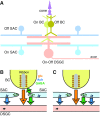
Figure 1.
Figure 1.
Models for On-Off DSGC synaptic inputs. A, Basic circuit for the On-Off DSGC. Bipolar cells (BCs) and SACs of both the On and Off subtypes synapse onto DSGC dendrites. B, Existing model of DS synapses. Three types of synaptic input onto a rightward-preferring (→) DSGC dendrite show DS. A rightward projecting SAC dendrite prefers rightward motion and releases acetylcholine (Ach; this dendrite would release GABA onto a leftward-preferring DSGC, which is not shown). A leftward projecting SAC dendrite prefers leftward motion and releases GABA. This SAC also releases GABA onto a bipolar terminal, inhibiting its glutamate (glu) release during leftward motion and consequently generating a rightward preference for glutamate release. Each synaptic connection (large colored arrow) has a direction preference indicated by a small black arrow. C, Proposed model of DS synapses. A leftward projecting SAC dendrite prefers leftward motion and releases GABA. Both leftward and rightward projecting SAC dendrites release Ach onto the DSGC so that the net input lacks tuning. Ach input from the rightward projecting SAC dendrite could be explained if Ach, but not GABA, acted over a relatively longer distance (i.e., spillover) without direct synaptic contact (Briggman et al., 2011; Vaney et al., 2012). The glutamate synapse lacks DS; no SAC input to the bipolar terminal is required, and any such input would lack net tuning. NP, No direction preference.
Figure 2.
Figure 2.
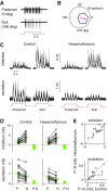
Tuning of mouse On-Off DSGC excitatory conductance persists with nicotinic receptors blocked. A, Spike response to motion in a DSGC's preferred and null direction. B, Polar plot showing spike rate, averaged over 5 s, to the grating moving in eight directions (same cell as in A). Red arrow indicates the vector mean angle of the response (preferred direction). C, Excitatory and inhibitory conductances for preferred- and null-direction motion in the control condition and in the presence of hexamethonium (100 μm). D, Synaptic conductance calculated for excitation (bottom) and inhibition (top) for responses, averaged over 5 s, to preferred (P) and null (N) directions. Mean values (±SEM) for P and N directions and the difference (PN) shown in green (n = 16 cells). E, The PN difference remained constant in the presence of hexamethonium (i.e., points lie near the identity line).
Figure 3.
Figure 3.
iGluSnFR signals on DSGC dendrites lack directional tuning. A, Two-photon images show selective, Cre-dependent expression of iGluSnFR in the CART-Cre retina. The image plane was at the On-layer dendrites of a DSGC filled with Alexa Fluor 594 (magenta). Box indicates the region in B1. B1, Fluorescence signals from a selected ROI (combination of subregions outlined with dashed lines) over trials. B2, Same as B1 for Off-layer ROI. C, Polar plot of the spike response for cell in A and B. D, Top, Average iGluSnFR signals for the ROI in B1 lacked directional tuning. Response was quantified by the peak-to-peak amplitude (F1:4 fit; red line). Bottom, Average excitatory conductance measured simultaneously with the imaging in B1 showed apparent DS. E, Same as D for the ROI in B2. F, Excitatory conductance was larger for preferred-direction motion compared with null-direction motion (i.e., points below the identity line). Recordings during imaging of On (magenta) and Off (black) layer dendrites are shown separately. Average response is shown in green. Error bars for individual points indicate ±SD across trials; error bars for the mean indicate ±SEM of the population (25 ROIs across n = 14 cells). G, Same as F for iGluSnFR signal amplitudes. Amplitudes were similar in the preferred and null directions.
Figure 4.
Figure 4.
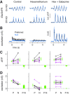
Apparent tuning in excitation can be explained by inhibition of the DSGC. A, iGluSnFR signals, averaged over four trials, in On and Off ROIs in a control condition and after adding hexamethonium (100 μm) and gabazine (50 μm). B, Excitatory conductance measured simultaneously with the iGluSnFR signals in A. C, iGluSnFR signal (peak-to-peak amplitude of F1:4 fit) for On (magenta) and Off (black) ROIs in response to preferred (P) and null (N) directions in each condition. Mean values (±SEM) for P and N directions and the difference (PN), shown in green (7–9 ROIs in each condition, across n = 4 cells). The iGluSnFR response lacked directional tuning in each condition. D, Same as C for excitatory conductance (response averaged over 5 s) measured simultaneously with iGluSnFR signals in C. The excitatory conductance showed apparent directional tuning in the control condition and in the presence of hexamethonium, but lacked tuning in the presence of gabazine.

References

    1. Barlow HB, Levick WR. The mechanism of directionally selective units in rabbit's retina. J Physiol. 1965;178:477–504. - PMC - PubMed
    1. Beier KT, Borghuis BG, El-Danaf RN, Huberman AD, Demb JB, Cepko CL. Transsynaptic tracing with vesicular stomatitis virus reveals novel retinal circuitry. J Neurosci. 2013;33:35–51. doi: 10.1523/JNEUROSCI.0245-12.2013. - DOI - PMC - PubMed
    1. Borghuis BG, Tian L, Xu Y, Nikonov SS, Vardi N, Zemelman BV, Looger LL. Imaging light responses of targeted neuron populations in the rodent retina. J Neurosci. 2011;31:2855–2867. doi: 10.1523/JNEUROSCI.6064-10.2011. - DOI - PMC - PubMed
    1. Borghuis BG, Marvin JS, Looger LL, Demb JB. Two-photon imaging of nonlinear glutamate release dynamics at bipolar cell synapses in the mouse retina. J Neurosci. 2013;33:10972–10985. doi: 10.1523/JNEUROSCI.1241-13.2013. - DOI - PMC - PubMed
    1. Brandon C. Cholinergic neurons in the rabbit retina: immunocytochemical localization, and relationship to GABAergic and cholinesterase-containing neurons. Brain Res. 1987;401:385–391. doi: 10.1016/0006-8993(87)91426-0. - DOI - PubMed

Publication types